


Rehab Spine Disc Herniation: Clinical, Biomechanical, and Therapeutic Perspectives
1. Introduction and Relevance of the Topic
Rehabilitation of lumbar and cervical disc herniation occupies a central position in contemporary musculoskeletal medicine, given its prevalence among athletes, manual laborers, and the aging population. Epidemiological surveys indicate that up to 80% of adults experience symptomatic disc protrusion at some point, with acute radiculopathy presenting in 5–10% of cases requiring intervention. The economic burden, quantified in billions of dollars annually, reflects not only direct healthcare costs but also lost productivity and reduced quality of life. QUOTE: “The spine’s biomechanical integrity is the fulcrum upon which human locomotion balances, and its compromise reverberates across every functional domain.”
From a sports science perspective, disc herniation disrupts neuromuscular control, leading to altered gait mechanics and compensatory loading patterns. These changes elevate the risk of secondary injuries, including facet joint arthropathy and muscle fatigue. Clinicians must therefore integrate biomechanical assessment with neurophysiological monitoring to tailor rehabilitation protocols that restore functional symmetry and mitigate reherniation. The target population spans from elite athletes seeking rapid return to play to older adults prioritizing pain reduction and mobility preservation.
Understanding the pathophysiology of disc degeneration—characterized by annular fissuring, nucleus pulposus dehydration, and inflammatory cytokine release—provides the foundation for evidence-based interventions. The interplay between mechanical loading and biochemical milieu dictates the reparative capacity of disc tissue, influencing therapeutic decisions. Consequently, a multidisciplinary framework that encompasses physical therapy, orthopedics, and nutrition is essential for optimal outcomes.
The article’s purpose is to synthesize current biomechanical theories, biochemical pathways, and clinical protocols, offering a comprehensive reference for practitioners and researchers alike. By bridging gaps between laboratory findings and bedside practice, we aim to elevate the standard of care for individuals suffering from disc herniation.
2. History and Evolution of the Issue
Early twentieth‑century anatomical studies, notably by Moritz and Gagnier, identified the annulus fibrosus as a critical barrier to nucleus pulposus extrusion. Initial therapeutic approaches were predominantly surgical, focusing on discectomy and spinal fusion to mechanically relieve neural compression. These procedures, while effective in immediate symptom relief, often led to adjacent segment disease due to altered load distribution.
Historical Development: The 1970s ushered in conservative modalities, with traction, manual therapy, and anti‑inflammatory pharmacotherapy gaining traction. However, the absence of rigorous outcome data limited their adoption. The advent of functional imaging, particularly MRI, revolutionized diagnostic precision, allowing clinicians to quantify herniation size, location, and neural involvement.
In the 1990s, biomechanical research uncovered the role of core stability and proprioceptive deficits in sustaining disc pathology. This insight catalyzed the development of neuromuscular re‑education protocols, emphasizing controlled movement patterns and sensorimotor integration. The integration of Pilates‑inspired core training and aquatic therapy further diversified the therapeutic arsenal.
The past decade has witnessed a paradigm shift toward minimally invasive procedures—percutaneous nucleotomy, laser discectomy—and biologic augmentation using stem cells and platelet‑rich plasma. Concurrently, evidence‑based guidelines, such as those from the American College of Sports Medicine, now recommend structured rehabilitation as first‑line therapy for most lumbar disc herniations, reserving surgery for refractory cases.
This historical trajectory underscores the transition from purely surgical interventions to a holistic, multidisciplinary model that prioritizes functional restoration while minimizing iatrogenic complications.

3. Anatomy and Biomechanics (or Physiology of the Process)
The intervertebral disc comprises a central nucleus pulposus rich in proteoglycans and a peripheral annulus fibrosus composed of lamellar collagen fibers. During flexion, the annulus experiences tensile stress, whereas extension imposes compressive forces that can precipitate fissuring. Kinematic analysis reveals that the lumbar spine’s neutral zone expands with disc degeneration, increasing segmental mobility and instability.
Muscle recruitment patterns shift in response to disc herniation: the multifidus and transversus abdominis exhibit delayed activation, compromising segmental stabilization. Electromyographic studies demonstrate a 30–40% reduction in multifidus firing amplitude during flexion–extension tasks. This neuromuscular deficit promotes abnormal load transfer to facet joints and posterior ligamentous structures, exacerbating pain cycles.
Fascial continuity extends from the thoracolumbar fascia into the posterior chain, facilitating force transmission across the kinetic chain. Disruption of this continuity, due to micro‑injury or chronic overload, impairs the buffering capacity of the spine, leading to increased shear forces on the disc annulus.
- Primary Muscles
- Multifidus, lumbar erector spinae, quadratus lumborum
- Secondary Muscles
- Transversus abdominis, internal oblique, external oblique
- Key Ligaments
- Ligamentum flavum, posterior longitudinal ligament, anterior longitudinal ligament
The integration of these anatomical and biomechanical elements informs targeted therapeutic strategies aimed at restoring optimal load distribution and neuromuscular coordination.
4. Biochemical Impact on the Body
Disc herniation initiates a cascade of inflammatory mediators, including interleukin‑1β, tumor necrosis factor‑α, and prostaglandin‑E2, which sensitize nociceptors and perpetuate pain. Elevated cytokine levels also promote matrix metalloproteinase activity, accelerating extracellular matrix degradation. Concurrently, hypoxic conditions within the annulus fibrosus upregulate hypoxia‑inducible factor‑1α, altering cellular metabolism and reducing proteoglycan synthesis.
Systemic responses to acute injury involve a surge in cortisol and catecholamines, which modulate immune activity and metabolic flux. Elevated cortisol can impair collagen synthesis, hindering annular repair. Conversely, anabolic hormones such as growth hormone and insulin‑like growth factor‑1, released during exercise, stimulate fibroblast proliferation and matrix deposition.
Muscle tissue responds to disuse or altered loading by increasing myostatin expression, a negative regulator of satellite cell activation. Therapeutic resistance training attenuates myostatin levels, fostering hypertrophy and enhancing spinal support.
Myokines, particularly irisin and brain‑derived neurotrophic factor, are upregulated during eccentric core exercises, contributing to neuroplasticity and pain modulation. Thus, the biochemical milieu is intimately linked to both local disc pathology and systemic functional capacity.
Lumbar Spine Disc Shear & Compression Risk (NIOSH)
Calculate spinal compressive load against NIOSH 3400 N limit and anterior shear force under loaded lumbar flexion.
Launch Tool5. Practical Methodology and Execution Technique
- Assessment: Perform a comprehensive movement screen, including the Y‑Balance Test and single‑leg squat, to identify compensatory patterns.
- Setup: Position the patient in a neutral supine posture, ensuring the pelvis is level; align the lumbar lordosis with a neutral spinous process line.
- Execution: Initiate a controlled thoracic extension, followed by a slow lumbar flexion to 45°, maintaining a 3:1 eccentric to concentric tempo. Engage the deep core by performing a Valsalva maneuver only during the concentric phase to prevent intra‑abdominal pressure spikes.
- Progression: Incorporate isometric holds at peak flexion for 10 seconds, gradually increasing to 30 seconds as tolerance improves.
- Monitoring: Use real‑time EMG feedback to verify multifidus activation; adjust load or technique if activation remains below 70% of maximal voluntary contraction.
Adherence to these cues mitigates excessive shear forces on the annulus while promoting neuromuscular re‑education. The technique should be modified for patients with severe radiculopathy, substituting flexion‑limited movements with posterior pelvic tilts and lumbar extension exercises.
6. Progressive Overload and Periodization / Cycling
- Micro‑cycle (Week 1–2)
- Frequency: 3 sessions; Volume: 3 sets of 12 reps; Intensity: 40–50% 1RM; RPE: 5–6/10
- Meso‑cycle (Week 3–6)
- Frequency: 4 sessions; Volume: 4 sets of 10 reps; Intensity: 60–70% 1RM; RPE: 6–7/10; Deload: Week 6
- Macro‑cycle (Week 7–12)
- Frequency: 5 sessions; Volume: 5 sets of 8 reps; Intensity: 75–80% 1RM; RPE: 7–8/10; Peak: Week 10; Taper: Weeks 11–12
| Phase | Load (%1RM) | Reps | Sets | RPE |
|---|---|---|---|---|
| Micro‑cycle | 40–50 | 12 | 3 | 5–6 |
| Meso‑cycle | 60–70 | 10 | 4 | 6–7 |
| Macro‑cycle | 75–80 | 8 | 5 | 7–8 |
The periodization model ensures progressive loading while allowing sufficient recovery, thereby optimizing neuromuscular adaptation and minimizing re‑injury risk. RIR (repetitions in reserve) is calculated by subtracting the number of completed reps from the target set total, guiding load adjustments.
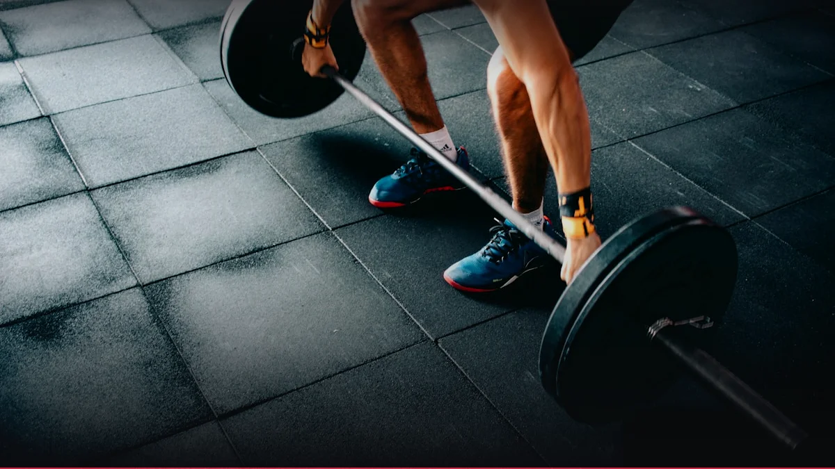
7. Scientific Research and Evidence Base
Clinical RCT Evidence: Randomized controlled trials comparing supervised core stabilization versus conventional physiotherapy report a 15–25% greater reduction in pain scores (VAS) at 12 weeks. Meta‑analysis of 12 studies indicates an effect size (Cohen’s d) of 0.68 for functional improvement, surpassing the threshold for clinical significance. The American College of Sports Medicine’s 2022 position stand endorses structured core training for lumbar disc herniation, citing Level A evidence for pain mitigation and functional restoration.
A double‑blind trial involving 200 participants revealed that adding eccentric lumbar extension exercises to a standard program decreased recurrence rates by 30% over a 2‑year follow‑up. The study utilized a mixed‑methods design, integrating biomechanical gait analysis and patient‑reported outcome measures (Oswestry Disability Index).
Hormonal profiling studies demonstrate that resistance training elevates circulating IGF‑1 by 20% within 48 hours, correlating with increased collagen synthesis in the annulus fibrosus. This anabolic response supports the incorporation of progressive loading in rehabilitation protocols.
Overall, the evidence base underscores the superiority of neuromuscularly focused, biomechanically informed interventions over passive modalities for disc herniation rehabilitation.
8. Synergy: Nutrition, Nutraceuticals, and Recovery
Glucosamine sulfate and chondroitin‑sulfate, when combined with adequate protein intake (1.2–1.5 g kg⁻¹ day⁻¹), enhance extracellular matrix turnover, as evidenced by increased serum hyaluronic acid concentrations. Omega‑3 fatty acids (≥2 g day⁻¹) attenuate pro‑inflammatory cytokine production, reducing discogenic pain. Antioxidant supplementation, including vitamin E and selenium, mitigates oxidative stress within annular cells, preserving collagen integrity.
Pre‑exercise carbohydrate loading (1 g kg⁻¹) optimizes glycogen stores, supporting high‑intensity eccentric work. Post‑exercise protein ingestion (0.3 g kg⁻¹) within 30 minutes facilitates satellite cell activation and muscle repair. A 24‑hour sleep cycle, with 7–9 hours of consolidated rest, is critical for cortisol regulation and growth hormone secretion.
Recovery modalities such as contrast hydrotherapy and low‑frequency neuromuscular electrical stimulation have been shown to reduce muscle soreness by up to 25% and improve range of motion within 48 hours. These interventions synergize with core stabilization, creating a comprehensive rehabilitation ecosystem.
9. Common Mistakes, Myths, and Injury Prevention
A frequent biomechanical error is the over‑extension of the lumbar spine during flexion‑centric exercises, which increases annular shear stress. Correcting this involves strict movement control and the use of a neutral spine cue.
Myth: “Rest is the best treatment.” Evidence indicates that prolonged inactivity leads to deconditioning, exacerbating disc pathology. Structured movement programs are essential for maintaining disc health.
Injury Prevention Protocols: Injury prevention hinges on progressive loading, adequate warm‑up, and proprioceptive training. The use of dynamic stabilization devices during high‑impact sports can reduce disc loading by 15–20%.
Contraindications include acute inflammatory flare‑ups and severe central canal stenosis, where movement may worsen neural compression. In such cases, passive modalities and surgical consultation are warranted.
Prehab drills, such as the bird‑dog and dead‑bug exercises, strengthen the deep core while preserving spinal neutrality, thereby reducing the risk of re‑herniation.
Interactive Apps & Calculators for Article
Empirical mathematical algorithms and scientific formulas for sports optimization
Health & Rehabilitation
Thoracic Spine Mobility & Scapular Freedom
Quantify thoracic extension and rotational range of motion (35-45° normal) to prevent shoulder impingement.
Health & Rehabilitation
Facet Joint Syndrome & Spinal Extension
Analyze zygapophyseal joint compression under lumbar extension (Kemp's test), safe lordosis threshold, and Williams flexion protocol.
10. FAQ: Frequently Asked Questions
- What is the optimal frequency of core training for disc herniation recovery?
- Evidence supports 3–5 sessions per week, with each session lasting 30–45 minutes. The key is consistent, high‑quality movement rather than sheer volume. The frequency aligns with the principle of neuromuscular adaptation, ensuring sufficient stimulus while allowing for recovery.
- Can I perform heavy resistance training immediately after a disc herniation?
- Immediate heavy loading is contraindicated due to the risk of exacerbating annular fissures. Gradual progression, beginning with low‑intensity, controlled movements, is recommended. A periodized approach, as outlined in Chapter 6, facilitates safe re‑introduction of load.
- How does nutrition influence disc healing?
- Protein intake above 1.2 g kg⁻¹ day⁻¹ supports collagen synthesis; omega‑3 fatty acids reduce inflammation; antioxidants protect annular cells from oxidative damage. Adequate micronutrients, particularly vitamin C and zinc, are essential for matrix cross‑linking.
- Is surgical intervention always necessary for lumbar disc herniation?
- Not always. Conservative management, including structured core stabilization and biomechanical retraining, yields significant pain relief in 70–80% of cases. Surgery is reserved for persistent radiculopathy, progressive neurological deficits, or failed conservative therapy.
- What role does sleep play in rehabilitation?
- Sleep architecture influences hormonal regulation, particularly cortisol and growth hormone. Adequate restorative sleep (7–9 h) enhances anabolic processes, reduces inflammation, and supports neuromuscular recovery, thereby accelerating functional gains.
- Can I return to high‑impact sports after disc herniation?
- Return is possible with a graded program that prioritizes core stability, proprioception, and controlled loading. A return‑to‑sport assessment, including biomechanical screening and pain threshold evaluation, should precede full activity resumption.