


Rehabilitation of the Runner's Knee: A Comprehensive Biomechanical and Physiological Analysis of Patellofemoral Pain Syndrome
1. Introduction and Relevance of the Topic
Patellofemoral pain syndrome, historically and colloquially referred to as "runner's knee," represents one of the most prevalent musculoskeletal injuries in the field of recreational and competitive endurance running. Epidemiological studies consistently indicate that approximately thirty to fifty percent of all running-related injuries involve the patellofemoral joint, making it a dominant cause of training interruption and long-term disability among athletes. The condition is characterized by anterior knee pain that is exacerbated by activities involving flexion under load, such as running downhill, ascending stairs, or prolonged sitting. The high incidence rate underscores the critical need for a nuanced understanding of the complex interplay between biomechanical inefficiencies, neuromuscular control deficits, and tissue tolerance thresholds that converge to produce this multifactorial pathology.
The clinical significance of patellofemoral pain syndrome extends beyond immediate pain management to encompass the preservation of joint integrity and the restoration of functional capacity. Unlike acute traumatic events, this syndrome often develops insidiously, driven by cumulative microtrauma and repetitive stress on the articular cartilage and subchondral bone. Consequently, effective rehabilitation requires a paradigm shift from passive, symptom-focused interventions to active, movement-based strategies that address the underlying etiological factors. This approach not only alleviates pain but also enhances the biomechanical resilience of the lower kinetic chain, thereby reducing the risk of recurrence and improving overall athletic performance and quality of life for affected individuals.
This article aims to provide an exhaustive, scientifically rigorous examination of the pathophysiology, biomechanics, and evidence-based rehabilitation protocols for patellofemoral pain syndrome. By integrating insights from orthopedic biomechanics, exercise physiology, and clinical rehabilitation science, we seek to delineate the precise mechanisms of injury and recovery. The content is structured to serve as a comprehensive reference for sports scientists, physiotherapists, and coaches, offering detailed protocols for assessment, intervention, and return-to-sport criteria. Understanding the intricate relationship between quadriceps force vectors, patellar tracking, and soft tissue compliance is essential for developing targeted, individualized treatment plans that yield sustainable long-term outcomes.
"The knee is not a solitary joint but a complex linkage system where dysfunction at any point in the kinetic chain can manifest as patellofemoral pain, necessitating a holistic rather than isolated approach to rehabilitation."
2. History and Evolution of the Issue
The historical understanding of patellofemoral pain syndrome has undergone a significant transformation from early mechanical theories to contemporary biomechanical and neuromuscular models. In the early twentieth century, clinicians primarily attributed anterior knee pain to static anatomical anomalies, such as patella alta or trochlear dysplasia, leading to interventions focused on surgical correction of bony malalignments. This reductionist view overlooked the dynamic nature of joint loading and the critical role of muscular control. As imaging technologies advanced, particularly with the introduction of magnetic resonance imaging, clinicians began to recognize that many individuals with significant anatomical variances remained asymptomatic, prompting a re-evaluation of the primary etiologies of pain.
The mid-twentieth century saw the emergence of the "maltracking" hypothesis, which posited that abnormal lateral displacement of the patella during knee extension was the central driver of patellofemoral stress. This model led to widespread adoption of therapeutic modalities aimed at correcting perceived lateral pull, including the use of taping, bracing, and specific stretching routines for the iliotibial band. However, these interventions often yielded inconsistent results, highlighting the limitations of a purely mechanical perspective. The failure to account for neuromuscular timing and strength deficits revealed a gap in the prevailing clinical dogma, suggesting that anatomical structure alone was insufficient to explain the variability in symptom onset and severity.
In recent decades, the paradigm has shifted decisively toward a multifactorial model that integrates biomechanical, physiological, and psychosocial factors. Modern research emphasizes the significance of poor neuromuscular control of the hip and knee, particularly during dynamic tasks like running. The concept of "dynamic malalignment" has replaced static geometric concerns, focusing on the real-time interaction between muscle forces and joint reaction forces. This evolution has led to evidence-based rehabilitation protocols that prioritize strengthening the hip abductors and external rotators, as well as the quadriceps, to optimize patellar tracking and load distribution, marking a definitive departure from earlier, less effective treatment strategies.

3. Anatomy and Biomechanics (or Physiology of the Process)
The patellofemoral joint is a sesamoid joint where the posterior surface of the patella articulates with the trochlear groove of the femur. This unique configuration allows the patella to increase the moment arm of the quadriceps tendon, thereby enhancing the mechanical advantage of knee extension. However, this efficiency comes at the cost of high contact pressures, particularly in the lower quarter of the patella during flexion beyond ninety degrees. The stress distribution is heavily influenced by the geometry of the trochlear groove and the patellar shape, with shallow grooves leading to increased lateral compression and shear forces. Understanding these kinematic parameters is crucial for identifying individuals at higher risk for cartilage degradation and pain.
The primary extrinsic stabilizers of the patella include the quadriceps femoris group, particularly the vastus medialis obliquus (VMO). The VMO plays a pivotal role in controlling patellar tracking by generating a medial force vector that counteracts the lateral pull of the vastus lateralis. The angle of pull of the VMO, known as the Q-angle, is a critical determinant of lateral patellar displacement. An increased Q-angle, often due to pelvic width or femoral anteversion, exacerbates lateral stress on the patellofemoral articulation. Neuromuscular timing is equally important, as delayed activation of the VMO during the terminal phases of knee extension can result in unopposed lateral force vectors, leading to maltracking and subsequent pain.
The kinematics of the patellofemoral joint are further modulated by the soft tissue envelope, including the retinaculum and the iliotibial band. While the ITB is not a direct stabilizer of the patella, its tension can indirectly influence patellar positioning through its attachment to the lateral femoral epicondyle and the lateral retinaculum. Excessive tightness or hypertonicity in the ITB can increase lateral compression forces, particularly during the mid-stance phase of running. The interplay between bony geometry, muscular force vectors, and soft tissue constraints creates a complex biomechanical environment where minor deviations in alignment or muscle activation can lead to significant increases in joint stress, precipitating the development of patellofemoral pain syndrome.
- Q-Angle (Quadriceps Angle)
- The angle formed by a line drawn from the anterior superior iliac spine to the center of the patella and a line from the patella to the tibial tuberosity. In females, the average Q-angle is approximately 17 degrees, compared to 14 degrees in males, contributing to higher lateral patellar stress.
- Patellar Tracking
- The path of the patella within the trochlear groove during knee flexion and extension. Normal tracking involves a smooth, centered glide, whereas maltracking involves excessive lateral displacement or tilting, increasing contact pressure on the lateral facet.
- Moment Arm
- The perpendicular distance from the axis of rotation to the line of action of a force. In the knee, the moment arm of the quadriceps determines the torque generated for extension; a larger moment arm allows for greater torque with less muscle force, but can also increase joint reaction forces.
4. Biochemical Impact on the Body
The pathophysiology of patellofemoral pain syndrome involves complex biochemical cascades triggered by mechanical overload and repetitive microtrauma. Increased contact pressures within the joint stimulate mechanoreceptors in the synovium and cartilage, leading to the release of pro-inflammatory cytokines such as interleukin-1 (IL-1) and tumor necrosis factor-alpha (TNF-alpha). These mediators initiate an inflammatory response, resulting in synovial hypertrophy and increased intra-articular pressure. The chronic presence of these inflammatory markers can downregulate collagen synthesis and upregulate matrix metalloproteinases (MMPs), leading to cartilage degradation and subchondral bone remodeling, which are hallmarks of early osteoarthritis.
Neuromuscular fatigue plays a critical role in exacerbating the biochemical environment of the injured knee. As the quadriceps and hip musculature fatigue, the ability to generate stabilizing force vectors diminishes, leading to increased reliance on passive structures and higher peak joint loads. This mechanical inefficiency further amplifies the mechanical stimulus for inflammation, creating a vicious cycle of pain and dysfunction. Additionally, pain itself alters motor control strategies, leading to compensatory movements that may increase stress on adjacent tissues. The central nervous system modulates pain perception through descending inhibitory pathways, which can become sensitized in chronic cases, leading to central sensitization and pain amplification even in the absence of peripheral pathology.
Rehabilitation interventions aim to reverse these negative biochemical adaptations by promoting an anti-inflammatory environment and enhancing tissue repair. Exercise-induced myokines, such as myostatin inhibitors and anti-inflammatory cytokines, are released during physical activity, helping to modulate the local inflammatory response. Furthermore, adequate nutrition and hydration support the biochemical processes necessary for tissue repair, including collagen cross-linking and glycosaminoglycan synthesis. The timing and intensity of exercise are crucial, as moderate loading stimulates anabolic pathways, while excessive loading can perpetuate catabolic states. Understanding these biochemical dynamics is essential for designing rehabilitation programs that optimize tissue healing and long-term joint health.
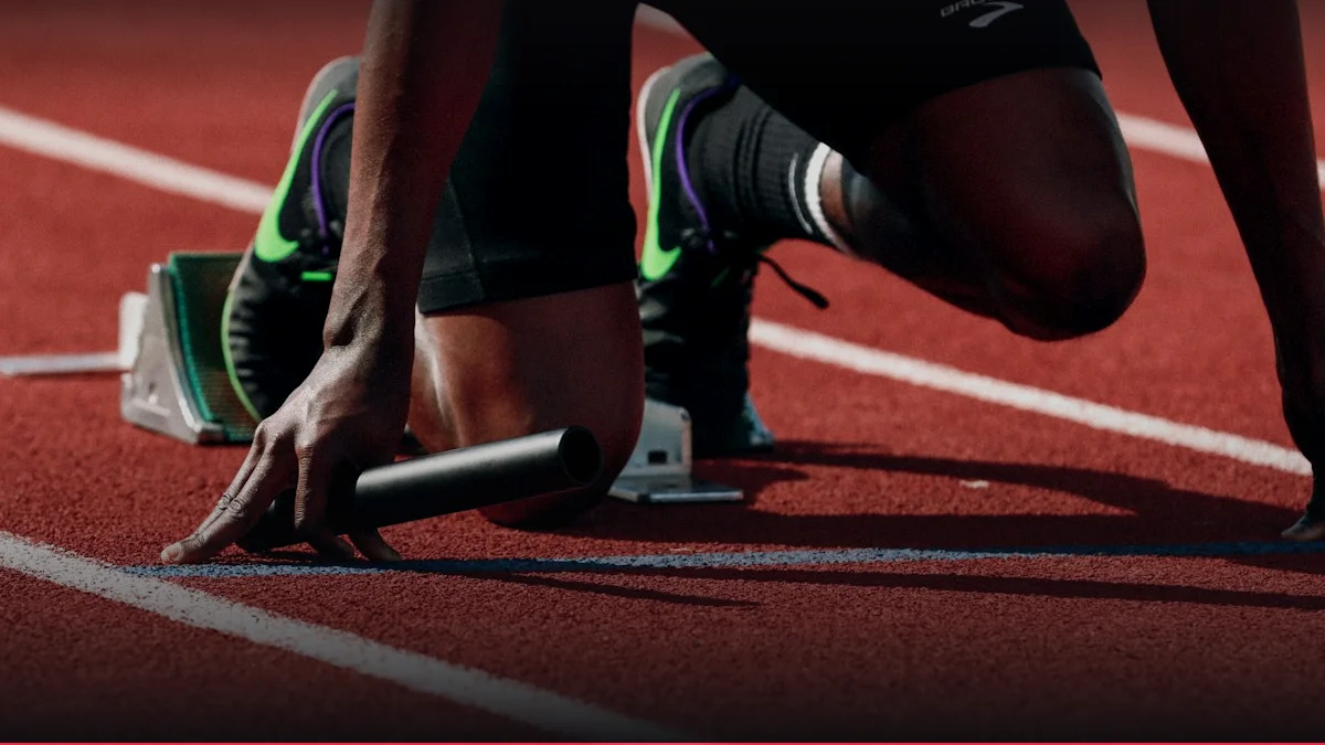
Runner's Knee Patellofemoral Stress & Cadence Fix
Calculate patellofemoral joint reaction forces and evaluate how increasing cadence by 7.5-10% reduces joint stress.
Launch Tool5. Practical Methodology and Execution Technique
Effective rehabilitation of patellofemoral pain syndrome requires a structured, progressive approach that addresses both neuromuscular control and strength deficits. The initial phase focuses on pain modulation and the re-establishment of optimal movement patterns. Key exercises include isometric quadriceps contractions, which allow for muscle activation without significant joint loading, and hip abductor strengthening to improve pelvic stability during single-leg activities. Proper execution is critical, with emphasis on maintaining neutral pelvic alignment and avoiding compensatory lateral trunk shifts.
The technique for performing terminal knee extension exercises involves starting from a fully extended position and actively contracting the quadriceps to lift the knee slightly off the surface, focusing on the medial aspect of the patella. This movement should be controlled and slow, ensuring that the patella glides smoothly within the trochlear groove. Breathing mechanics are also important, with exhalation during the concentric phase to maintain intra-abdominal pressure and core stability. Patients should be cued to focus on the sensation of the VMO contracting, which can be facilitated by placing a hand on the medial aspect of the quadriceps to provide tactile feedback.
As pain levels decrease and strength improves, the rehabilitation program progresses to dynamic, functional movements that mimic the demands of running. This includes single-leg squats, step-downs, and lateral band walks, all performed with strict attention to hip and knee alignment. The cue "knees over toes" may be used to ensure proper femoral tracking, while "push the knees out" helps engage the hip abductors. Tempo is crucial, with a focus on controlled eccentric loading to strengthen the musculature and improve tissue tolerance. Gradual reintroduction to running should follow a structured progression, increasing volume and intensity only when the patient can perform daily activities and strength exercises without pain.
- Position the patient in a supine position with the affected leg extended.
- Instruct the patient to tighten the thigh muscles and press the back of the knee into the floor.
- Maintain the contraction for five to ten seconds while exhaling slowly.
- Release the tension and relax the leg for a few seconds before repeating.
- Perform three sets of ten repetitions, ensuring no pain is experienced during the exercise.
6. Progressive Overload and Periodization / Cycling
The design of a rehabilitation program for patellofemoral pain syndrome must adhere to the principles of progressive overload and periodization to ensure sustainable improvements in strength, endurance, and function. The macro-cycle typically spans several months, divided into mesocycles that target specific physiological adaptations. The initial mesocycle focuses on pain reduction and neuromuscular activation, utilizing low-intensity, high-frequency exercises. As the patient progresses, the mesocycle shifts toward strength development, increasing the resistance and complexity of movements. This structured approach prevents overtraining and allows for adequate recovery, which is essential for tissue repair and adaptation.
Micro-cycling within each mesocycle involves the careful management of training volume and intensity to avoid exacerbating symptoms. The use of Rate of Perceived Exertion (RPE) and Repetitions in Reserve (RIR) provides objective metrics for monitoring workload. For example, a patient might perform sets of ten to fifteen repetitions at an RPE of five to seven, ensuring that the last few repetitions can be completed with good form. Deload weeks are incorporated every three to four weeks to allow for supercompensation and to reduce the risk of overuse injuries. This cyclical approach ensures that the body is constantly adapting to new stimuli, leading to continuous improvements in biomechanical efficiency and tissue tolerance.
The table below summarizes the key parameters for each phase of the rehabilitation periodization, providing a clear framework for clinicians and athletes to follow. Each phase has specific goals, exercise types, and progression criteria that must be met before advancing to the next stage. This systematic approach ensures that the rehabilitation process is both safe and effective, maximizing the chances of a successful return to sport.
| Phase | Duration | Primary Goal | Exercise Intensity | Progression Criteria |
|---|---|---|---|---|
| Phase 1: Pain Modulation | 2-4 Weeks | Reduce pain, activate muscles | Low RPE (3-5), Isometric | Pain-free daily activities |
| Phase 2: Strength Building | 4-6 Weeks | Increase quadriceps/hip strength | Moderate RPE (6-7), Dynamic | Single-leg squat without pain |
| Phase 3: Functional Training | 4-6 Weeks | Improve running mechanics | High RPE (8-9), Plyometric | Run 20 mins pain-free |
| Phase 4: Return to Sport | 2-4 Weeks | Full athletic performance | Maximal RPE (10), Sport-specific | Full training load tolerated |
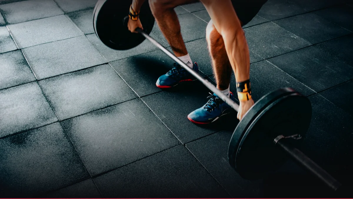
7. Scientific Research and Evidence Base
A substantial body of peer-reviewed literature supports the efficacy of strengthening exercises in the treatment of patellofemoral pain syndrome. Randomized controlled trials have consistently demonstrated that programs focusing on hip and quadriceps strength lead to significant reductions in pain and disability compared to traditional care or sham interventions. Meta-analyses indicate that strengthening alone is as effective as, or more effective than, combining strengthening with other modalities such as taping or bracing. The effect sizes for pain reduction are generally large, suggesting that these interventions have a clinically meaningful impact on patient outcomes.
The mechanism of action for these strengthening programs is thought to involve improved neuromuscular control and reduced joint loading. By increasing the strength and endurance of the hip abductors and external rotators, athletes can maintain better pelvic stability during single-leg support, thereby reducing the medial genu valgum and lateral patellar displacement. Similarly, strengthening the quadriceps, particularly the VMO, enhances the ability to control patellar tracking and distribute joint stresses more evenly. These biomechanical improvements translate into lower peak contact pressures, which is the primary driver of pain in patellofemoral pain syndrome.
ACSM Consensus: The American College of Sports Medicine (ACSM) and the International Society of Arthroplasty (ISSN) position stands endorse the use of exercise-based rehabilitation as the first-line treatment for patellofemoral pain syndrome. These guidelines emphasize the importance of individualized programs that address specific deficits identified through clinical assessment. They also highlight the need for patient education and self-management strategies to promote long-term adherence and prevent recurrence. The consensus is that a multi-faceted approach, combining strength training, flexibility, and education, yields the best outcomes for this common running injury.
8. Synergy: Nutrition, Nutraceuticals, and Recovery
Nutrition plays a pivotal role in the rehabilitation of patellofemoral pain syndrome by supporting tissue repair, modulating inflammation, and optimizing energy availability. Adequate protein intake is essential for muscle protein synthesis and the repair of damaged soft tissues. The timing of protein consumption, particularly in the post-exercise window, can enhance recovery and adaptation. Additionally, the inclusion of anti-inflammatory nutrients, such as omega-3 fatty acids and antioxidants, can help mitigate the inflammatory response associated with repetitive joint loading. A balanced diet that supports overall metabolic health is crucial for achieving optimal recovery outcomes.
Hydration status also significantly impacts joint health and muscle function. Dehydration can reduce synovial fluid volume, increasing friction and stress within the patellofemoral joint. It can also impair muscle contractility and neuromuscular coordination, leading to poorer movement quality and increased injury risk. Therefore, maintaining proper hydration before, during, and after exercise is a critical component of any rehabilitation program. Electrolyte balance is also important, as imbalances can lead to muscle cramps and fatigue, further compromising biomechanical efficiency.
Sleep is another critical factor in recovery, as it is during this time that the body releases growth hormone and other anabolic factors necessary for tissue repair. Poor sleep quality or insufficient sleep duration has been linked to increased pain sensitivity and slower recovery from exercise. Therefore, promoting good sleep hygiene and ensuring adequate rest are essential for optimizing the recovery process. The synergy between nutrition, hydration, and sleep creates a favorable internal environment for healing, enhancing the effectiveness of physical therapy and exercise interventions.
9. Common Mistakes, Myths, and Injury Prevention
One of the most common mistakes in the management of patellofemoral pain syndrome is the reliance on passive modalities such as ice, ultrasound, and manual therapy as standalone treatments. While these interventions may provide temporary symptom relief, they do not address the underlying biomechanical and neuromuscular deficits that cause the condition. A focus on active rehabilitation is essential for long-term success. Another common error is the premature return to high-intensity running without adequate strength and conditioning, which can lead to symptom recurrence and chronicity.
Myths surrounding the condition, such as the belief that running is inherently harmful to the knee or that stretching the ITB is the primary solution, persist despite evidence to the contrary. Research shows that running is generally safe for healthy knees and that the ITB is not a primary contributor to patellofemoral pain. Instead, the focus should be on strengthening the entire kinetic chain and improving movement quality. Educating patients about these myths is crucial for managing expectations and ensuring adherence to evidence-based protocols.
Injury Prevention Protocols: Injury prevention strategies should focus on building resilience and improving biomechanical efficiency. This includes incorporating strength training into regular training routines, ensuring proper warm-up and cool-down, and gradually increasing training load. Cross-training with low-impact activities can also help maintain fitness while reducing joint stress. Regular screening for neuromuscular deficits and early intervention for pain or discomfort can prevent minor issues from developing into chronic problems. A proactive approach to knee health is essential for endurance athletes aiming to maintain long-term performance and health.
Interactive Apps & Calculators for Article
Empirical mathematical algorithms and scientific formulas for sports optimization
Health & Rehabilitation
Meniscus Knee Compressive Load & Shear Angle
Quantify tibiofemoral compressive loads and posterior horn shear forces across 0° to 135° deep knee flexion angles.
Health & Rehabilitation
Knee Sleeves vs Wraps: Rebound Kinetic Energy & Patellar Load
Compare 7mm neoprene sleeves (+5-10 kg) versus 2.5m powerlifting wraps (+20-35 kg): elastic stored energy (Joules) and patellofemoral compressive stress.
10. FAQ: Frequently Asked Questions
- Does running cause patellofemoral pain syndrome?
- Running itself does not cause patellofemoral pain syndrome; rather, the condition is often a result of how the running mechanics interact with individual biomechanical factors. Repetitive loading can exacerbate pre-existing weaknesses or imbalances, leading to pain. However, once the underlying issues are addressed through strengthening and technique modification, running is generally safe and beneficial for joint health.
- How long does rehabilitation typically take?
- The duration of rehabilitation varies depending on the severity of the condition and the individual's response to treatment. Most patients can expect to see significant improvements within six to twelve weeks of consistent, evidence-based exercise. Full return to sport-specific activities may take several months, requiring a gradual progression of load and intensity to ensure long-term success.
- Are braces or taping effective for long-term management?
- Braces and taping can provide temporary relief by altering patellar tracking and reducing pain during activity. However, they are not a standalone solution for long-term management. They should be used as adjuncts to a comprehensive rehabilitation program that addresses the underlying neuromuscular deficits. Relying solely on external support can lead to muscle deconditioning and dependence.
- What is the role of the VMO in patellofemoral pain?
- The vastus medialis obliquus (VMO) is a key muscle for controlling patellar tracking. Weakness or delayed activation of the VMO can lead to excessive lateral displacement of the patella, increasing contact pressure and pain. Strengthening the VMO, often through specific isolation exercises, is a critical component of rehabilitation to restore balanced force vectors around the knee joint.
- Can I continue to run while I have pain?
- It is generally recommended to modify activity levels to avoid exacerbating pain. Low-impact cross-training can be used to maintain fitness while the knee heals. Once pain levels decrease, a gradual return to running can be initiated, following a structured progression. Listening to the body and adjusting intensity based on pain response is crucial to prevent flare-ups and ensure a safe recovery.
- Is surgery ever necessary for patellofemoral pain syndrome?
- Surgery is rarely indicated for patellofemoral pain syndrome and is typically reserved for cases with significant structural abnormalities or failure of conservative treatment. Most cases respond well to non-surgical interventions, including exercise, education, and activity modification. Surgical options, such as tibial tubercle transfer, carry risks and do not always guarantee a successful outcome, so they are considered a last resort.