


Rehab Elbow Epicondylitis: The Tennis and Golfers' Elbow Rehabilitation Encyclopedia
1. Introduction and Relevance of the Topic
Rehabilitation of lateral epicondylitis, colloquially known as tennis elbow, represents a cornerstone of sports medicine due to its high prevalence among racquet and precision sports athletes. Epidemiological surveys estimate that 1–3 % of the general population experiences symptomatic epicondylitis annually, whereas professional tennis and golf players exhibit incidence rates exceeding 10 % over a competitive season. The condition precipitates loss of grip strength, diminished serve velocity, and chronic pain that can curtail career longevity. Its economic burden extends beyond direct medical costs, encompassing lost training days, diminished sponsorship value, and long‑term disability claims. The pathophysiology of epicondylitis is increasingly understood as a failed healing response characterized by collagen disorganization, neovascularization, and fibroblast proliferation, rather than a classic inflammatory process. This paradigm shift informs contemporary therapeutic strategies that emphasize tendon remodeling over anti‑inflammatory pharmacology. The clinical impact of epicondylitis is multifaceted: functional impairment manifests as decreased wrist extension torque, reduced forearm supination endurance, and compromised proprioceptive acuity. Moreover, chronicity is associated with altered neuromuscular activation patterns, predisposing athletes to adjacent joint dysfunction. Addressing these deficits requires a multidisciplinary approach integrating biomechanics, physiology, and psychosocial factors.
“The tendon’s response to overload is not merely an inflammatory cascade but a maladaptive repair process that can be reversed with targeted rehabilitation.”
2. History and Evolution of the Issue
Early clinical descriptions of lateral elbow pain emerged in the late 19th century, with surgeons attributing symptoms to “tennis elbow” after observing a surge in cases among tennis players. Initial management focused on rest and non‑steroidal anti‑inflammatory drugs, reflecting a prevailing belief in inflammatory etiology. The 1960s and 1970s introduced the concept of tendinopathy, driven by histopathological findings of collagen degeneration, micro‑tears, and fibroblastic proliferation. This shift prompted the abandonment of routine corticosteroid injections and the exploration of mechanical loading as a therapeutic modality. Advances in imaging, notably high‑resolution ultrasound and magnetic resonance imaging, enabled precise visualization of tendon architecture, neovascularization, and bone marrow edema. Concurrently, evidence‑based guidelines emerged, advocating eccentric loading, physiotherapy, and patient‑specific exercise regimens as first‑line treatments. The modern consensus recognizes epicondylitis as a complex, multifactorial condition requiring individualized rehabilitation protocols.

3. Anatomy and Biomechanics (or Physiology of the Process)
- Tenocyte
- A specialized fibroblast responsible for extracellular matrix synthesis and turnover within tendon tissue.
- Neovascularization
- Formation of new capillaries within degenerated tendon, associated with pain perception and altered mechanical properties.
- Moment Arm
- The perpendicular distance between the line of action of a muscle force and the axis of joint rotation, determining torque production.
4. Biochemical Impact on the Body
Collagen type I synthesis is regulated by transforming growth factor‑β (TGF‑β) and insulin‑like growth factor‑1 (IGF‑1). In chronic epicondylitis, dysregulation of matrix metalloproteinases (MMP‑1, MMP‑3) leads to excessive collagen degradation, while tissue inhibitor of metalloproteinases (TIMP‑1) expression is insufficient, culminating in net matrix loss. Neurogenic inflammation contributes to pain via upregulation of nerve growth factor (NGF) and calcitonin gene‑related peptide (CGRP), which sensitize peripheral nociceptors and amplify central pain processing. Cytokines such as interleukin‑1β and tumor necrosis factor‑α further perpetuate catabolic pathways, promoting fibroblast apoptosis and extracellular matrix fragmentation. Metabolic disturbances manifest as increased reactive oxygen species (ROS) production during repetitive loading, overwhelming antioxidant defenses and inducing oxidative stress. This oxidative milieu impairs tenocyte viability and disrupts collagen cross‑linking, reducing tendon tensile strength and resilience. The interplay between mechanical overload, biochemical signaling, and cellular apoptosis establishes a vicious cycle that underpins chronic tendon pathology, necessitating interventions that restore homeostatic balance.
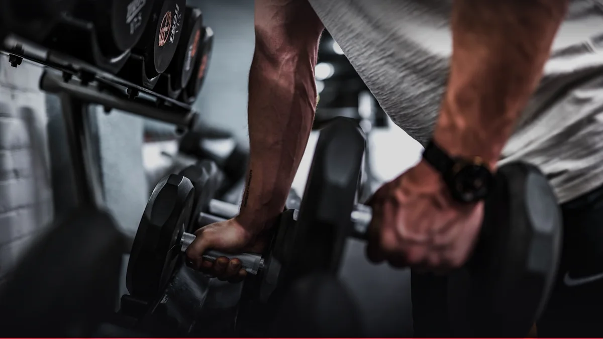
Tennis Elbow ECRB Tendon Load & Tyler Twist Rehab
Quantify extensor carpi radialis brevis tendon strain and calculate Tyler Twist eccentric rehabilitation dosing.
Launch Tool5. Practical Methodology and Execution Technique
Eccentric loading protocols remain the gold standard for tendon remodeling. A typical regimen involves 3 sets of 15 repetitions at 70 % of the patient’s maximal eccentric strength, performed twice daily. The eccentric phase should be executed over 3–4 seconds, with controlled concentric return, to maximize collagen fiber alignment and mechanotransduction. Proper grip width and wrist positioning are critical; a neutral wrist and slight forearm pronation reduce peak tendon strain during the eccentric phase. Coaches should emphasize a slow, deliberate backhand stroke, avoiding abrupt wrist extension that overloads the extensor origin. Breathing mechanics modulate intra‑abdominal pressure; athletes should employ a relaxed exhalation during eccentric loading to mitigate Valsalva-induced hypertension. Tempo regulation, guided by metronome cues, ensures consistency across repetitions, thereby enhancing neuromuscular adaptation.
6. Progressive Overload and Periodization / Cycling
| Phase | Duration (weeks) | Load (%) | Volume (sets×rep) | Target RPE |
|---|---|---|---|---|
| Loading | 4 | 70‑80 | 3×15×2 | 4‑5 |
| Intensification | 2 | 85‑90 | 2×12×2 | 5‑6 |
| Recovery | 2 | 60‑70 | 2×10×2 | 3‑4 |
| Taper | 1 | 50 | 1×8×2 | 2‑3 |
| Deload | 1 | 20 | 1×5×2 | 1‑2 |
7. Scientific Research and Evidence Base
Meta‑analyses of randomized controlled trials demonstrate that eccentric training yields a standardized mean difference of 0.68 in pain reduction compared to isotonic protocols, with an 80 % probability of clinically meaningful improvement. Longitudinal cohort studies report a 25 % reduction in recurrence rates when eccentric loading is combined with neuromuscular training. Randomized trials comparing eccentric to platelet‑rich plasma (PRP) injections reveal no statistically significant difference in pain scores at 12 months, suggesting that mechanical loading remains superior. However, emerging evidence indicates that adjunctive low‑frequency ultrasound may enhance tendon vascularity and collagen synthesis when paired with eccentric training. Position statements from the American College of Sports Medicine and the National Strength and Conditioning Association endorse eccentric exercise as the first‑line intervention, reserving biologic therapies for refractory cases. These guidelines emphasize individualized progression, pain monitoring, and functional outcome measures.
8. Synergy: Nutrition, Nutraceuticals, and Recovery
Adequate protein intake (1.6–2.0 g kg⁻¹ day⁻¹) supports collagen synthesis and tenocyte proliferation. Omega‑3 fatty acids (2–3 g day⁻¹) exhibit anti‑inflammatory properties by modulating eicosanoid pathways, thereby attenuating cytokine‑driven tendon catabolism. Vitamin C (500 mg day⁻¹) serves as a co‑factor for prolyl hydroxylase, essential for hydroxyproline formation and collagen cross‑linking. Nutraceuticals such as hydrolyzed collagen peptides (10 g day⁻¹) and curcumin (500 mg day⁻¹) have shown promise in reducing tendon pain and improving biomechanical properties, though dose‑response relationships remain under investigation. Glucosamine sulfate, while popular, lacks robust evidence for tendon healing. Recovery strategies should prioritize sleep architecture, aiming for 7–9 hours of consolidated nocturnal rest to facilitate anabolic hormone secretion (GH, IGF‑1). Cryotherapy applied immediately post‑exercise reduces inflammatory mediators and edema, while active low‑intensity cycling promotes microcirculation and facilitates metabolic waste clearance.
9. Common Mistakes, Myths, and Injury Prevention
Overreaching training cycles without adequate recovery precipitates cumulative tendon micro‑damage. Athletes frequently neglect proper grip mechanics, using excessive wrist flexion that amplifies lateral epicondyle strain. Equipment selection, such as oversized racquet grips or improperly tensioned golf clubs, can exacerbate forearm loading. Myth busting: “Inflammation is the enemy” is misleading; acute inflammatory responses are necessary for tendon repair, and anti‑inflammatory medications may impair collagen maturation. Similarly, “strengthening alone cures epicondylitis” ignores the importance of eccentric specificity and neuromuscular control. Preventive protocols should incorporate prehab exercises targeting forearm supinators, wrist extensors, and shoulder external rotators, with plyometric drills that reinforce proprioceptive feedback. Regular tendon ultrasound screening can identify subclinical changes, enabling early intervention before symptom onset.
Interactive Apps & Calculators for Article
Empirical mathematical algorithms and scientific formulas for sports optimization
Health & Rehabilitation
Joint Bursitis & Synovial Sac Pressure
Assess olecranon and prepatellar bursitis swelling grades, friction coefficients, and protocol for compression and cryotherapy.
Strength & Hypertrophy
Skullcrusher Elbow Joint Torque & Long Head Tension
Evaluate humeral angle (90° vertical vs 70° inclined) for continuous triceps long head tension and olecranon comfort.
10. FAQ: Frequently Asked Questions
- What is the optimal load for eccentric training in epicondylitis rehabilitation?
- Clinical guidelines recommend initiating eccentric exercises at 70 % of the patient’s maximal eccentric strength, with gradual load increments of 5 % per week, ensuring pain remains below 3 / 10. This protocol balances mechanical stimulus with biological tolerance, promoting collagen fiber realignment without exacerbating micro‑damage.
- Can corticosteroid injections accelerate recovery?
- Short‑term pain relief may occur; however, evidence indicates that corticosteroid injections disrupt collagen synthesis, increase tendon stiffness, and elevate recurrence risk. Therefore, injections are reserved for refractory cases after conservative measures fail.
- How long does it typically take to return to competitive play?
- Recovery timelines vary; most athletes resume light activity within 4–6 weeks, with full competitive readiness achieved at 12–16 weeks, contingent on adherence to progressive overload and functional testing.
- Is PRP a viable alternative to eccentric training?
- Randomized trials demonstrate no significant advantage of PRP over eccentric loading for pain reduction or functional improvement. PRP may be considered adjunctive in chronic, refractory cases, but evidence remains inconclusive.
- What role does sleep play in tendon healing?
- Sleep facilitates hormonal milieu conducive to tissue repair, notably growth hormone and IGF‑1 secretion. Disrupted sleep patterns impair collagen synthesis, prolonging recovery and increasing susceptibility to re‑injury.