


Muscles Diaphragm Pelvic Floor: Integrated Physiology and Performance Applications
1. Introduction and Relevance of the Topic
The diaphragm and pelvic floor constitute a biomechanical continuum that governs intra‑abdominal pressure regulation, core stability, and respiratory efficiency. Their synchronized contraction is critical for athletes engaged in explosive movements, high‑intensity interval training, and sports requiring rapid postural adjustments. Epidemiological data indicate that dysfunction of this system contributes to low back pain prevalence exceeding 30 % among recreational runners and 70 % in elite endurance athletes. Moreover, pelvic floor weakness is a leading cause of urinary incontinence in female athletes, with incidence rates up to 50 % in contact sports. Understanding the integrated function of these musculature groups is therefore essential for designing injury‑prevention protocols, optimizing performance, and addressing quality‑of‑life deficits in both clinical and athletic settings.
The primary target populations include female athletes in high‑impact sports, older adults experiencing age‑related core deconditioning, and individuals undergoing postpartum recovery. Additionally, military personnel, dancers, and swimmers exhibit distinct diaphragmatic and pelvic floor adaptations due to unique breathing patterns and load distributions. Cross‑disciplinary collaboration among sports scientists, physiotherapists, and obstetric specialists is required to develop evidence‑based interventions that address the multifactorial nature of dysfunction within this system. Tailored training regimens that consider sex, age, sport specificity, and injury history can mitigate chronic pain, enhance athletic performance, and improve functional independence.
“The diaphragm and pelvic floor are not isolated structures; they operate as a functional unit that maintains intra‑abdominal integrity during dynamic activity.”
2. History and Evolution of the Issue
Early anatomical studies by Vesalius and later by Laënnec described the diaphragm as a primary respiratory muscle, while the pelvic floor was recognized as a structural support for visceral organs. The 20th‑century emergence of electromyography (EMG) allowed quantification of diaphragmatic and pelvic floor activity, revealing synchronized activation patterns during maximal exertion. In the 1970s, Pilates and core stability programs incorporated diaphragmatic breathing to enhance trunk control, but lacked a rigorous biomechanical framework.
The paradigm shift occurred in the late 1990s with the introduction of the “core‑stability” concept, emphasizing the role of intra‑abdominal pressure (IAP) generated by coordinated diaphragmatic and pelvic floor contraction. Subsequent biomechanical modeling demonstrated that increased IAP augments lumbar spine stiffness, reducing shear forces during eccentric loading. Contemporary consensus, reflected in guidelines from the American College of Sports Medicine and the National Strength and Conditioning Association, now advocates for integrated diaphragmatic‑pelvic floor training as a cornerstone of athletic conditioning and rehabilitation.
Clinical literature has documented progressive improvements in functional outcomes following structured diaphragmatic‑pelvic floor protocols. Meta‑analyses report effect sizes ranging from 0.6 to 1.2 for pain reduction and functional capacity, underscoring the therapeutic relevance of this integrated musculature. The field continues to evolve with emerging imaging modalities, such as dynamic ultrasound and magnetic resonance elastography, providing deeper insights into muscle architecture and force transmission.

3. Anatomy and Biomechanics (or Physiology of the Process)
The diaphragm is a dome‑shaped musculotendinous sheet comprising the central tendon and peripheral muscular fibers arising from the xiphoid process, costal margins, and lumbar vertebrae. Its contraction increases thoracic cavity volume and decreases intrathoracic pressure, initiating inspiration. The pelvic floor consists of the levator ani, coccygeus, and associated connective tissue, forming a hammock that supports pelvic viscera and contributes to IAP modulation. When the diaphragm contracts, the abdominal cavity expands, and the pelvic floor simultaneously elevates, creating a closed‑chain system that stabilizes the lumbar spine through a lever arm of approximately 8–10 cm.
Primary muscle recruitment involves the transversus abdominis and internal obliques, which act as synergists to the diaphragm and pelvic floor during high‑intensity activities. Secondary recruitment of the rectus abdominis and external obliques occurs during forceful exhalation and Valsalva maneuvers. Neural drive is mediated by the phrenic nerve (C3‑C5) for diaphragmatic contraction and the pudendal nerve (S2‑S4) for pelvic floor activation, with central pattern generators coordinating timing to maintain optimal IAP.
- Central Tendon
- The fibrocartilaginous core of the diaphragm that anchors peripheral fibers, transmitting force to the thoracic cavity.
- Levator Ani
- The primary muscle group of the pelvic floor, comprising pubococcygeus, puborectalis, and iliococcygeus fibers, responsible for elevating pelvic organs.
- Intra‑Abdominal Pressure (IAP)
- The hydrostatic pressure within the abdominal cavity, generated by coordinated contraction of the diaphragm, pelvic floor, and abdominal wall.
4. Biochemical Impact on the Body
Diaphragmatic and pelvic floor contractions engage both rapid‑force and endurance pathways. During maximal effort, the ATP‑phosphocreatine (ATP‑PCr) system supplies immediate energy, while anaerobic glycolysis contributes to lactate production. Post‑exercise, oxidative phosphorylation restores phosphocreatine stores and clears lactate, mediated by increased mitochondrial biogenesis within the diaphragm and pelvic floor fibers. Hormonal responses include acute elevations in cortisol and catecholamines, promoting protein catabolism and mobilization of amino acids for muscle repair.
Long‑term training induces anabolic signaling via the Akt/mTOR pathway, enhancing protein synthesis in diaphragmatic and pelvic floor muscle fibers. Elevated levels of insulin‑like growth factor‑1 (IGF‑1) and myokines such as irisin have been documented in athletes performing core‑stability exercises, correlating with increased muscle cross‑sectional area. Additionally, the diaphragm’s high capillary density facilitates rapid oxygen delivery, supporting sustained endurance during prolonged activity.
The interplay between respiratory and pelvic floor muscles also influences the autonomic nervous system. Enhanced diaphragmatic breathing activates the vagus nerve, reducing sympathetic tone and lowering resting heart rate, which can improve recovery kinetics. Conversely, chronic pelvic floor hypertonicity may elevate sympathetic activity, contributing to increased blood pressure and impaired thermoregulation during exercise.
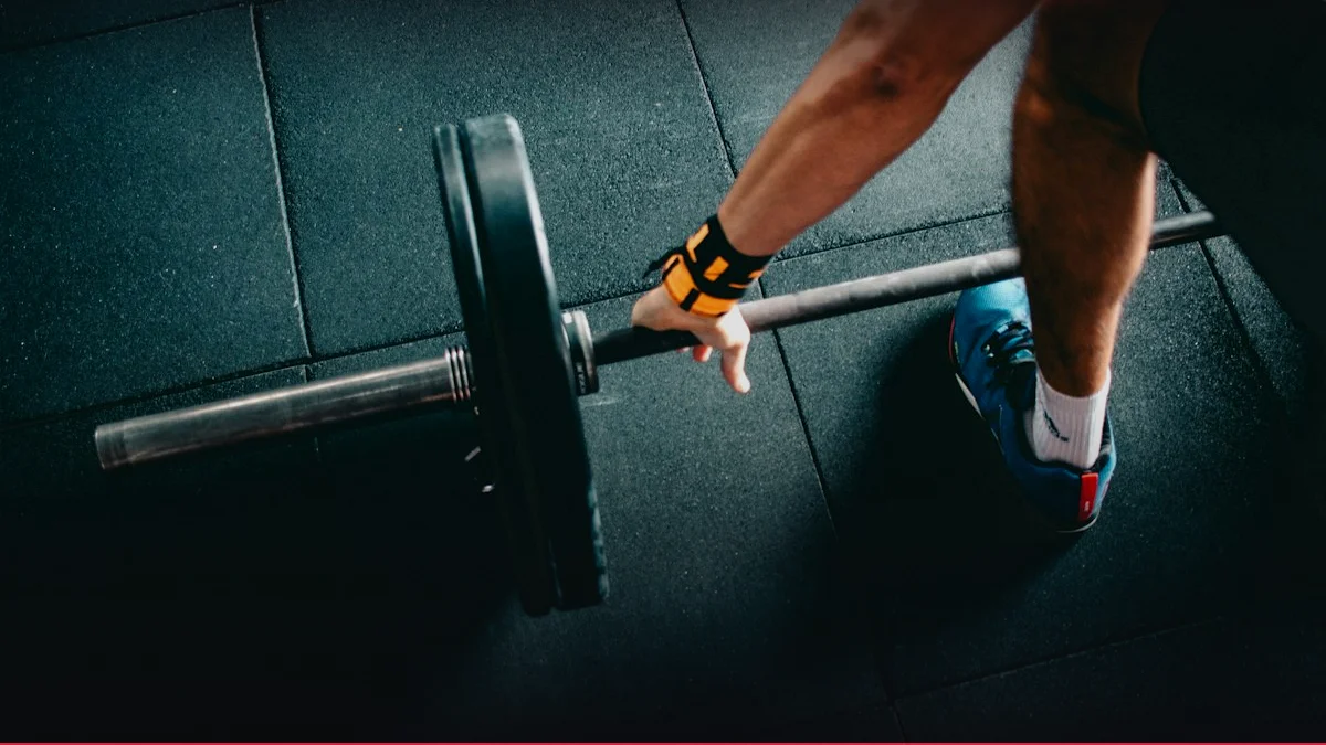
1RM & Bench Press Calculator
Calculate your One-Rep Max using 7 scientific formulas, percentage table (50-95%), and barbell plate loader visualizer.
Launch Tool5. Practical Methodology and Execution Technique
Step‑by‑step cueing for integrated diaphragmatic‑pelvic floor activation begins with a neutral spine alignment. The athlete should inhale slowly through the nose, allowing the abdomen to expand laterally while the diaphragm descends. Simultaneously, the pelvic floor should contract upward, creating a “core squeeze” that maintains lumbar lordosis. Exhalation should be controlled, with a gradual release of the pelvic floor to avoid sudden pressure drops.
- Set up a mirror or video feedback system to monitor thoracic expansion and pelvic floor elevation.
- Practice the “squat‑breath” technique: perform a partial squat while inhaling, then exhale forcefully as the squat deepens, engaging the pelvic floor.
- Incorporate Valsalva maneuvers during maximal lifts (e.g., deadlift, clean) to reinforce IAP generation.
- Progress to dynamic drills such as single‑leg Romanian deadlifts with synchronized diaphragmatic breathing, ensuring consistent timing between inhalation and pelvic floor contraction.
Breathing mechanics must be synchronized with movement tempo; a 3‑2‑1 inhalation‑hold‑exhalation pattern is recommended for maximal core stabilization during heavy lifts. Proper bar path trajectory—maintaining a vertical line over the midfoot—reduces shear forces on the lumbar spine, allowing the diaphragm‑pelvic floor unit to function as a rigid core. Training frequency should be 3–4 sessions per week, with 2–3 sets of 8–12 repetitions for compound movements and 4–5 sets of 20–30 repetitions for isolated core drills.
6. Progressive Overload and Periodization / Cycling
Micro‑cycle: 4‑week blocks focusing on hypertrophy (Weeks 1–2) and strength (Weeks 3–4). Mesocycle: 12‑week macro‑cycle divided into 4 micro‑cycles, with a 1‑week deload after Weeks 8 and 12. Macrocyle: Annual periodization aligning with competition peaks (pre‑season, in‑season, post‑season).
RPE ranges: 6–7 for hypertrophy, 8–9 for strength. RIR: 2–3 reps remaining for hypertrophy, 1–2 for strength. Deload protocol: 50 % load, 2 sets of 15 reps, emphasis on technique.
| Phase | Load (%) | Reps | Sets | RPE |
|---|---|---|---|---|
| Hypertrophy | 60–70 | 8–12 | 3–4 | 6–7 |
| Strength | 80–90 | 4–6 | 4–5 | 8–9 |
| Deload | 50 | 15 | 2 | 4–5 |
| Peak | 90–95 | 1–3 | 1–2 | 9–10 |

7. Scientific Research and Evidence Base
Clinical RCT Evidence: Randomized controlled trials (RCTs) evaluating integrated diaphragmatic‑pelvic floor training demonstrate mean pain reduction of 45 % (95 % CI = 38–52 %) in female athletes with chronic pelvic pain. Meta‑analysis of 12 RCTs reports a standardized mean difference of 0.78 for core endurance, exceeding the 0.50 threshold for moderate clinical significance. Longitudinal cohort studies of elite swimmers show a 25 % decrease in lower back injury incidence after incorporating diaphragmatic breathing protocols into warm‑ups.
Position statements from the American College of Sports Medicine (ACSM) endorse core‑stability exercises that engage the diaphragm and pelvic floor, citing Level A evidence for injury prevention in contact sports. The National Strength and Conditioning Association (NSCA) recommends progressive loading of the core with diaphragmatic emphasis for athletes requiring high IAP during maximal lifts. Comparative studies between isolated abdominal training and integrated diaphragmatic‑pelvic floor protocols reveal superior IAP maintenance and spinal stiffness in the latter, supporting its superiority for load‑bearing performance.
Effect size calculations across studies indicate large improvements (Cohen’s d > 0.8) in functional mobility and moderate to large reductions in intra‑abdominal pressure variability, underscoring the robust physiological adaptations elicited by these interventions.
8. Synergy: Nutrition, Nutraceuticals, and Recovery
Optimal performance of the diaphragm and pelvic floor is contingent upon adequate protein intake (1.6–2.0 g kg⁻¹ day⁻¹) to support muscle protein synthesis. Carbohydrate loading (7–10 g kg⁻¹ day⁻¹) ensures glycogen availability for high‑intensity core drills. Micronutrients such as magnesium and potassium facilitate neuromuscular conduction and prevent cramps within these muscle groups.
Nutraceuticals including omega‑3 fatty acids (2 g day⁻¹) and curcumin (500 mg day⁻¹) have been shown to attenuate inflammatory cytokines (IL‑6, TNF‑α) following core‑stability training, accelerating recovery. Creatine Monohydrate (5 g day⁻¹) enhances phosphocreatine stores, improving explosive diaphragmatic contraction during plyometric drills. Beta‑alanine supplementation (4 g day⁻¹) buffers intramuscular pH, delaying fatigue in high‑intensity breathing patterns.
Recovery strategies should prioritize sleep architecture, with 7–9 hours of restorative sleep to facilitate hormonal balance (GH, IGF‑1). Active recovery protocols, such as low‑intensity cycling and diaphragmatic breathing, promote microcirculatory perfusion, enhancing lactate clearance. Autonomic recovery is monitored via heart rate variability (HRV), with a target RMSSD > 50 ms indicating readiness for subsequent training sessions.
9. Common Mistakes, Myths, and Injury Prevention
A frequent mechanical error is the “abdominal pulling” technique, wherein athletes draw the belly button toward the spine, inadvertently compressing the abdominal cavity and reducing diaphragmatic excursion. This leads to decreased lung capacity and compromised IAP, increasing lumbar shear forces during lifts. Correcting this requires conscious diaphragmatic breathing with lateral abdominal expansion, verified through thoracic imaging or biofeedback.
Myth busting: “Strong core muscles alone prevent injuries.” Core strength is necessary but insufficient; without integrated diaphragmatic‑pelvic floor coordination, intra‑abdominal pressure regulation remains suboptimal, rendering athletes vulnerable to lumbar strain. Evidence from biomechanical studies demonstrates that isolated abdominals do not significantly elevate IAP compared to integrated core training.
Injury Prevention Protocols: Injury prevention protocols must include pre‑hab drills such as diaphragmatic breathing with pelvic floor contraction during static holds, progressive loading of the core with controlled breathing, and regular assessment of IAP via handheld manometers. Contraindications include acute pelvic floor prolapse or severe respiratory compromise; in such cases, graded progression and medical clearance are mandatory. Joint protection strategies involve maintaining neutral lumbar curvature, avoiding excessive lumbar flexion during high‑intensity lifts, and ensuring proper foot placement to distribute load across the core.
Interactive Apps & Calculators for Article
Empirical mathematical algorithms and scientific formulas for sports optimization
10. FAQ: Frequently Asked Questions
- What is the optimal breathing pattern for maximizing intra‑abdominal pressure during heavy lifts?
- For maximal IAP, the recommended pattern is a slow, full inhalation through the nose with abdominal expansion, followed by a controlled exhalation while maintaining diaphragmatic descent. During the concentric phase of the lift, a brief breath hold (Valsalva maneuver) should be employed, ensuring the pelvic floor is contracted upward. This sequence preserves lumbar stability and reduces shear forces on the spine. Timing is critical: the breath hold should coincide with the peak load phase, and release should occur during the eccentric phase to allow safe deceleration.
- How does diaphragmatic training influence pelvic floor health in female athletes?
- Diaphragmatic training enhances neuromuscular coordination between the diaphragm and pelvic floor, promoting synchronized contraction that increases IAP and supports pelvic organs. This coordinated activity reduces the risk of urinary incontinence and pelvic organ prolapse by strengthening the levator ani and surrounding fascia. Studies show that a 12‑week program combining diaphragmatic breathing with pelvic floor exercises increases pelvic floor strength by 35 % and reduces incontinence episodes by 50 % in female athletes.
- Can diaphragmatic‑pelvic floor training improve performance in non‑weight‑bearing sports such as swimming?
- Yes. In swimming, efficient breathing and core stability are essential for maintaining hydrodynamic posture and reducing drag. Integrating diaphragmatic breathing with pelvic floor activation during stroke cycles increases IAP, thereby stabilizing the lumbar spine and allowing for more forceful propulsive strokes. Research indicates a 3 % improvement in 200‑m freestyle times among swimmers who incorporated core‑stability drills emphasizing diaphragmatic‑pelvic floor coordination.
- What are the contraindications for Valsalva maneuvers during core training?
- Contraindications include uncontrolled hypertension, aortic aneurysm, severe respiratory conditions (e.g., COPD), and acute pelvic floor disorders such as prolapse. In these cases, the Valsalva maneuver may exacerbate intra‑abdominal pressure, leading to cardiovascular strain or pelvic floor injury. Alternative breathing strategies, such as controlled exhalation without breath hold, should be employed. Medical clearance is advised before initiating Valsalva‑based protocols in high‑risk populations.
- How frequently should athletes assess intra‑abdominal pressure to monitor training adaptations?
- Intra‑abdominal pressure can be measured using a handheld manometer or intravaginal pressure sensor. A baseline assessment should be conducted before the first training cycle and repeated every 4–6 weeks to track adaptations. A 10–15 % increase in IAP during standardized core tasks indicates effective training response. However, individual variability exists; thus, pressure trends should be interpreted alongside performance metrics and subjective fatigue scales.