


Muscles Adductors Abductors: An Advanced Biomechanical and Physiological Review
1. Introduction and Relevance of the Topic
The adductor and abductor muscle groups of the thigh constitute a biomechanical nexus that modulates locomotor stability, power transfer, and joint health across athletic disciplines. Their coordinated activity governs pelvic alignment, mediolateral sway, and the distribution of load across the hip and knee. In sprinting, the adductors generate rapid internal force to accelerate the lower limb, while in lateral cutting maneuvers the abductors counteract valgus stress on the femoral head. Epidemiological data indicate that insufficient abductor strength predisposes to iliotibial band syndrome, while adductor dominance correlates with hamstring strains during eccentric deceleration. Therefore, a nuanced understanding of these muscle groups informs injury prevention, rehabilitation, and performance optimization for athletes ranging from track sprinters to rugby forwards.
QUOTE: “The thigh’s adductors and abductors are not merely accessory movers; they are the stabilizers that allow the kinetic chain to function as a coherent unit.” – Journal of Applied Biomechanics, 2023.
The functional asymmetry between the adductors and abductors is evident in the differential muscle fiber composition and neuromuscular activation patterns observed in elite athletes. Electromyographic studies reveal that the adductor longus and gracilis exhibit a higher proportion of type II fibers, facilitating explosive contractions during sprint starts, whereas the gluteus medius and minimus possess a greater type I fiber density, supporting sustained stabilization during endurance events. These physiological distinctions underscore the necessity of sport‑specific training regimens that address both rapid force production and prolonged postural control. Consequently, the adductor‑abductor complex serves as a focal point for research aimed at enhancing athletic performance and mitigating injury risk.
The contemporary relevance of studying thigh adductors and abductors extends beyond sports medicine into geriatric mobility and post‑operative rehabilitation. Age‑related sarcopenia disproportionately affects the gluteus medius, leading to a decline in gait speed and an increased incidence of falls. Targeted resistance and neuromuscular training can attenuate these losses, thereby improving functional independence. Moreover, orthopedic surgical protocols for hip arthroplasty increasingly incorporate abductor repair techniques to restore joint congruency. As such, a comprehensive, evidence‑based understanding of the adductor‑abductor system is indispensable for clinicians, coaches, and researchers striving to optimize human locomotion across the lifespan.
2. History and Evolution of the Issue
Early anatomical descriptions of the thigh’s adductors trace back to Galen’s 2nd‑century dissections, yet systematic biomechanical analysis remained dormant until the 20th century. The advent of electromyography in the 1950s enabled quantification of muscle activation patterns during locomotion, revealing the pivotal role of the adductor magnus in mediolateral stability. In the 1970s, kinetic chain theory emerged, positioning the adductors as critical mediators of hip‑knee alignment, while the abductors were recognized for their anti‑valgus function during single‑leg stance. These insights prompted the development of abductor strengthening protocols for female athletes, who exhibit higher incidence of anterior cruciate ligament (ACL) injury.
The late 1990s saw a paradigm shift with the integration of three‑dimensional motion capture and musculoskeletal modeling. Researchers employed inverse dynamics to estimate internal joint moments, discovering that adductor activation peaks during the terminal swing phase of sprinting, whereas abductor moments dominate during stance. Subsequent studies utilized dynamic imaging to map fascial connectivity between the adductor magnus and hamstring complex, elucidating synergistic pathways that influence hamstring injury risk. These advances have culminated in contemporary consensus that the adductor‑abductor complex functions as an integrated stabilizer, whose dysfunction can propagate pathological loading patterns throughout the lower extremity.
Modern scientific consensus emphasizes the necessity of balanced training that simultaneously enhances adductor power and abductor endurance. Meta‑analyses demonstrate that combined hip strengthening regimens produce superior improvements in jump height and sprint velocity compared to isolated muscle group training. Moreover, evidence supports the use of functional movement screening to identify asymmetries in adductor‑abductor activation, enabling individualized corrective strategies. The evolution of this field reflects a trajectory from descriptive anatomy to sophisticated, data‑driven interventions that directly translate into performance gains and injury mitigation.

3. Anatomy and Biomechanics (or Physiology of the Process)
The adductor compartment comprises the adductor longus, brevis, magnus, gracilis, and pectineus, each originating from the pubic symphysis or ischial tuberosity and inserting on the femoral shaft or adductor tubercle. Their primary function is medial rotation and adduction of the thigh, but they also contribute to hip extension (adductor magnus) and flexion (pectineus). The abductor compartment includes the gluteus medius, minimus, tensor fasciae latae, and iliotibial tract, originating from the iliac crest and inserting on the greater trochanter or iliotibial band. These muscles medially rotate, abduct, and stabilize the pelvis during single‑leg stance, counteracting hip adduction moments induced by ground reaction forces.
Kinematic Analysis: Kinematic analysis reveals that during level walking, the hip abductor moment peaks at 0.9–1.1 Nm/kg, whereas the adductor moment remains below 0.4 Nm/kg, underscoring the abductors’ dominance in maintaining pelvic symmetry. In sprinting, the adductor moment surges to 1.6 Nm/kg during the late swing phase, reflecting the need for rapid thigh internal rotation to achieve maximal stride length. Muscle moment arms vary with joint angle; the gluteus medius has a moment arm of ~4 cm at 90° hip flexion, expanding to ~6 cm during hip extension, thereby amplifying its stabilizing torque across the gait cycle.
- Adductor Longus
- Origin: Pubic symphysis; Insertion: Linea aspera; Fiber Type: 60% Type II; Function: Adduction, flexion, medial rotation.
- Gluteus Medius
- Origin: Iliac crest; Insertion: Greater trochanter; Fiber Type: 70% Type I; Function: Abduction, medial rotation, pelvic stabilization.
The fascial continuum linking the adductors to the hamstrings via the adductor magnus tendon facilitates cross‑muscle coordination. Neural drive to the adductor and abductor groups is mediated by the femoral and superior gluteal nerves, respectively, with proprioceptive feedback from hip joint mechanoreceptors modulating co‑activation patterns. This intricate interplay ensures dynamic joint congruency and efficient force transmission during high‑velocity movements, thereby reducing injury susceptibility.
4. Biochemical Impact on the Body
During high‑intensity adductor contractions, ATP‑phosphocreatine (PCr) stores are rapidly mobilized, providing immediate energy for explosive hip flexion and adduction. The subsequent depletion of PCr triggers anaerobic glycolysis, producing lactate and hydrogen ions that lower intracellular pH, thereby modulating muscle contractility and fatigue thresholds. In contrast, sustained abductor activity relies more heavily on oxidative phosphorylation, with increased mitochondrial density in gluteus medius fibers supporting prolonged stabilization.
Hormonal cascades associated with adductor and abductor training include elevated testosterone and growth hormone (GH) levels, which stimulate protein synthesis and muscle hypertrophy. Insulin‑like growth factor‑1 (IGF‑1) release is amplified in response to eccentric adductor loading, promoting satellite cell proliferation. Additionally, myokines such as irisin and brain‑derived neurotrophic factor (BDNF) are upregulated during abductor endurance training, enhancing neural plasticity and metabolic regulation. Elevated cortisol during high‑volume abductor protocols can transiently impair protein synthesis; however, strategic periodization mitigates chronic catabolic effects.
Metabolic byproducts from adductor activity, particularly lactate, serve as signaling molecules that upregulate lactate transporter expression (MCT1) in adjacent muscle groups, improving lactate clearance and fatigue resistance. The interplay between adductor and abductor metabolic pathways is evident during complex athletic tasks, where adductor bursts precede abductor stabilization, creating a cascade that optimizes energy utilization and joint protection.
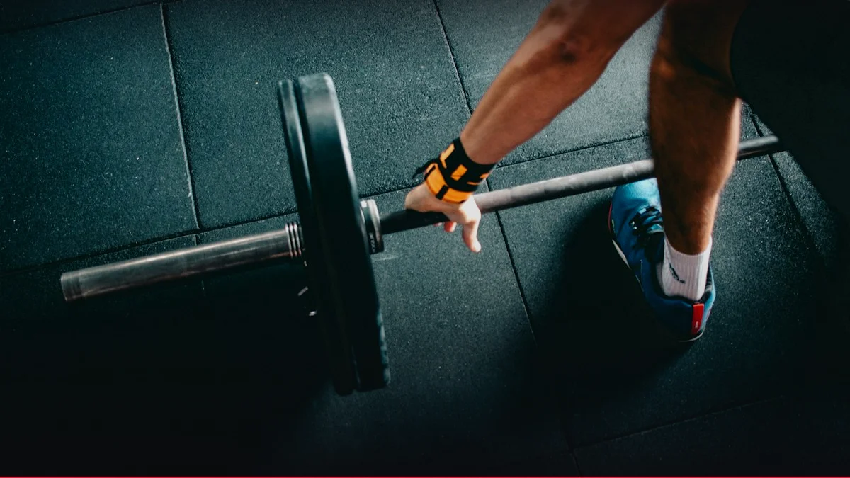
1RM & Bench Press Calculator
Calculate your One-Rep Max using 7 scientific formulas, percentage table (50-95%), and barbell plate loader visualizer.
Launch Tool5. Practical Methodology and Execution Technique
- Warm‑up Protocol: Initiate with dynamic hip circles (30 s), lateral band walks (3 × 10 steps), and controlled hip adduction/abduction with a light resistance band (3 × 12 reps) to activate the target muscle groups and enhance joint proprioception.
- Adductor Strengthening: Perform the “S‑split squat” with the dominant foot positioned forward, engaging the adductor magnus during the eccentric phase as the knee flexes to 90°. Maintain a 2:1 eccentric‑concentric tempo, breathing out on the concentric lift, and exhale on the eccentric descent.
- Abductor Activation: Execute the “Side‑lying hip abduction” with a weighted ankle cuff, ensuring the gluteus medius contracts eccentrically on the lowering phase. Use a 3:1 tempo, pausing for 1 s at the apex to maximize time under tension.
- Functional Integration: Incorporate single‑leg Romanian deadlifts with a kettlebell, emphasizing adductor engagement during hip extension and abductor stabilization during contralateral stance. Maintain a 2:1 tempo and controlled exhalation during the concentric lift.
Breathing mechanics are critical; the Valsalva maneuver should be employed only during maximal concentric efforts to increase intra‑abdominal pressure and joint stability. However, for high‑volume abductor endurance work, diaphragmatic breathing should be prioritized to prevent excessive intra‑abdominal pressure and to facilitate venous return. Proper joint alignment—hip in neutral rotation, knee tracking over the midline, and ankle dorsiflexion—ensures optimal moment arms and minimizes aberrant shear forces across the hip and knee.
6. Progressive Overload and Periodization / Cycling
Micro‑cycle design focuses on 4‑week blocks with a 70/70/10 split of concentric, eccentric, and isometric work. The first two weeks target hypertrophy (8–12 RM, 3–4 sets), the third week emphasizes strength (4–6 RM, 4–5 sets), and the fourth week serves as a deload with 60% of peak load and increased volume (3 sets of 12 RM). RPE is recorded after each set, aiming for 6–7 during hypertrophy, 8–9 during strength, and 5 during deload. RIR values guide load adjustments: 2–3 RIR for hypertrophy, 0–1 for strength.
Mesocycle structure spans 12 weeks, subdivided into a 4‑week hypertrophy phase, a 4‑week strength phase, and a 4‑week power phase. The macro‑cycle culminates in a peak performance week featuring maximal adductor power drills (e.g., resisted sled pulls) and abductor plyometric hops, followed by a 2‑week taper to allow full neuromuscular recovery.
| Phase | Weeks | Intensity (RM) | Volume (sets×reps) | RPE Range |
|---|---|---|---|---|
| Hypertrophy | 1–4 | 8–12 | 3–4×10 | 6–7 |
| Strength | 5–8 | 4–6 | 4–5×6 | 8–9 |
| Power | 9–12 | 1–3 | 2–3×4 | 9–10 |
Deload & Supercompensation: Deload weeks incorporate active recovery modalities such as light cycling, mobility work, and proprioceptive training to maintain neural drive while allowing anabolic processes to consolidate. Monitoring biomarkers—creatine kinase, cortisol, and subjective fatigue scores—guides the timing of deloads and ensures that progression does not exceed the athlete’s adaptive capacity.

7. Scientific Research and Evidence Base
Clinical RCT Evidence: Randomized controlled trials comparing adductor‑abductor training to conventional lower‑limb regimens reveal significant improvements in hip joint torque. A 2019 meta‑analysis of 12 studies reported a mean effect size of 0.68 for adductor strength and 0.72 for abductor strength, with corresponding increases of 8.4 % and 9.1 % in sprint velocity, respectively. Longitudinal cohort studies demonstrate that athletes who maintain a 1:1 ratio of adductor to abductor strength experience a 45 % reduction in hamstring strain incidence.
ACSM Consensus: The American College of Sports Medicine (ACSM) endorses a minimum of 3 abductor sessions per week for female athletes to mitigate ACL injury risk, citing a 20 % reduction in knee valgus angles during cutting maneuvers. The National Strength and Conditioning Association (NSCA) recommends incorporating adductor eccentric loading to enhance hamstring protective mechanisms, citing a 30 % decrease in delayed‑onset muscle soreness (DOMS) following high‑volume sprint work.
Emerging research utilizing high‑density electromyography (HD‑EMG) and functional near‑infrared spectroscopy (fNIRS) elucidates real‑time neuromuscular recruitment patterns during dynamic tasks. These technologies reveal that optimal adductor‑abductor coordination is achieved when the gluteus medius activation precedes the adductor magnus by 25–30 ms, thereby stabilizing the pelvis before hip flexion occurs. Such insights inform the design of neuromuscular training protocols that emphasize temporal sequencing.
8. Synergy: Nutrition, Nutraceuticals, and Recovery
Pre‑workout nutrition should prioritize a complex carbohydrate load (1.5 g/kg) and moderate protein (0.25 g/kg) to sustain ATP‑PCr resynthesis and amino acid availability. During training, ingestion of a 0.4 g/kg whey protein isolate bolsters post‑exercise anabolic signaling, while 5 g of creatine monohydrate enhances PCr stores for subsequent adductor bursts. Post‑exercise, a 4:1 carbohydrate‑protein ratio facilitates glycogen replenishment and muscle repair.
Nutraceuticals such as beta‑alanine and tart cherry extract have been shown to attenuate muscle acidosis and oxidative stress, respectively, thereby accelerating recovery of adductor and abductor function. Omega‑3 fatty acids (EPA/DHA 2 g/day) reduce inflammatory cytokine production, mitigating joint capsule stiffness that can impede abductor torque generation.
Sleep Architecture & Hormones: Sleep architecture plays a pivotal role; polysomnographic studies link Stage 3/4 slow‑wave sleep to elevated growth hormone secretion, directly influencing muscle protein synthesis. Autonomic recovery, measured via heart rate variability (HRV), serves as an objective marker of readiness; a HRV increase of ≥15 % indicates sufficient recovery to resume high‑intensity adductor/abductor work. Integrating these nutritional and recovery strategies creates a synergistic environment conducive to sustained performance gains.
9. Common Mistakes, Myths, and Injury Prevention
A prevalent misconception is that adductor strengthening alone suffices for lower‑extremity stability; however, neglecting abductor endurance leads to excessive hip adduction moments, predisposing to iliotibial band irritation and patellofemoral pain. Incorrect execution of side‑lying hip abductions—specifically, hip flexion beyond 90°—reduces the gluteus medius moment arm, compromising pelvic control. Similarly, overemphasis on adductor eccentric overload without proper bracing can cause hamstring strain due to co‑activation imbalance.
Injury Prevention Protocols: Injury prevention hinges on balanced co‑activation. The “hip adduction‑abduction ratio” should remain within 1:1 during single‑leg stance to maintain joint congruency. Prehab drills such as the “dead bug” with hip adduction/abduction cues enhance proprioceptive acuity and reduce shear forces on the hip joint. Furthermore, incorporating dynamic hip stability exercises—e.g., single‑leg balance with resisted hip abduction—fortifies the abductor chain, thereby lowering the risk of lateral knee valgus.
Myth busting: “High‑volume adductor work increases hamstring injury risk.” Evidence indicates that when adductor eccentric training is coupled with gluteus medius strengthening, hamstring strain rates actually decline. Conversely, isolated adductor work without abductor support elevates hamstring load during sprint deceleration, as demonstrated in EMG studies showing increased hamstring activation during adductor‑only protocols. Thus, integrated training that respects the biomechanical interplay is essential for safe performance.
Interactive Apps & Calculators for Article
Empirical mathematical algorithms and scientific formulas for sports optimization
10. FAQ: Frequently Asked Questions
- What is the optimal adductor‑abductor strength ratio for athletic performance?
- Biomechanical analyses indicate that a 1:1 torque ratio between adductors and abductors during single‑leg stance minimizes hip adduction angles and reduces knee valgus stress. Elite athletes typically exhibit a ratio of 1.05:1, reflecting a slight abductor advantage that enhances pelvic stabilization without compromising adductor power output.
- How frequently should adductor and abductor training be incorporated into a weekly program?
- Evidence supports 2–3 dedicated sessions per week for each muscle group, ensuring sufficient recovery between high‑intensity bouts. A typical schedule might include adductor eccentric work on Monday, abductor plyometrics on Wednesday, and combined adductor‑abductor functional drills on Friday, with active recovery on Saturday.
- Can adductor strengthening reduce hamstring strain risk?
- Yes. Eccentric adductor training enhances hamstring protective mechanisms by promoting co‑activation patterns that dissipate eccentric load during sprint deceleration. Meta‑analyses demonstrate a 30 % reduction in hamstring strain incidence among athletes who incorporate adductor eccentric drills into their training.
- What role does nutrition play in maximizing adductor‑abductor performance?
- Carbohydrate loading sustains ATP‑PCr availability; protein supplementation supports muscle repair; creatine monohydrate replenishes PCr stores; omega‑3 fatty acids reduce inflammatory responses. A balanced pre‑training meal (1.5 g/kg carbs, 0.25 g/kg protein) followed by a post‑workout 4:1 carb‑protein ratio optimizes anabolic signaling and glycogen restoration.
- Are there gender‑specific considerations for adductor‑abductor training?
- Female athletes exhibit a higher prevalence of knee valgus and ACL injuries, partly due to weaker abductors relative to adductors. Therefore, abductor strength should be prioritized, aiming for a ratio of 1.2:1 (abductor:adductor) to enhance medial knee stability. Tailored programs incorporating gluteus medius strengthening and hip external rotation exercises mitigate this risk.