


Iron and Anemia: Energy Deficit and Oxygen Starvation in Athleticism
1. Introduction and Relevance of the Topic
In the relentless pursuit of elite athletic performance, coaches and sports scientists frequently focus on complex periodization models, neuromuscular priming, and sports nutrition stacks. However, this hyper-specialized focus often overlooks a fundamental physiological reality: cellular respiration, mitochondrial ATP generation, and muscle fiber contraction depend entirely on adequate intracellular oxygen delivery. Iron deficiency is one of the most prevalent, silent metabolic bottlenecks in competitive sports, systematically degrading maximal aerobic capacity (VO2 max), impairing neuromuscular firing frequency, and accelerating peripheral fatigue across both endurance and strength disciplines. Recognizing this microscopic limitation is essential for sustained athletic longevity and performance progression.
Epidemiological investigations consistently reveal that up to 35% of female athletes and 15% to 20% of male endurance competitors suffer from subclinical iron depletion without exhibiting overt clinical anemia. This hidden deficiency manifests as progressive training stagnation, unexplained increases in resting heart rate, prolonged recovery windows, and early lactate accumulation during submaximal efforts. The physiological toll of compromised iron status extends far beyond hemoglobin-mediated oxygen transport, directly disrupting mitochondrial cristae bioenergetics and sarcomeric cross-bridge cycling speed.
QUOTE: Elite athletic potential remains fundamentally capped by the biochemical availability of intracellular oxygen transport mechanisms, rendering iron homeostasis the silent architect of sustainable performance ceilings.
Modern sports medicine requires a definitive paradigm shift from reactive treatment of severe clinical anemia toward proactive hematological monitoring and targeted iron optimization. By integrating periodized nutritional strategies with objective biochemical assessments, sports practitioners can eliminate the insidious metabolic bottlenecks that quietly undermine hard-won training adaptations. This comprehensive approach ensures that muscle hypertrophy, cardiovascular remodeling, and mitochondrial biogenesis advance in synchrony without encountering cellular oxygen starvation.
2. The Role of Iron in the Organism: Oxygen Transport and ATP Synthesis
Iron functions as an indispensable catalytic cofactor across dozens of enzymatic pathways that govern human bioenergetics, mitochondrial respiration, and physical exertion. Approximately 65% to 70% of total body iron resides within the heme moiety of hemoglobin molecules packed into circulating erythrocytes. Each hemoglobin tetramer contains four iron-protoporphyrin IX complexes that reversibly bind up to four oxygen molecules in pulmonary capillaries. This oxygenated complex traverses the cardiovascular tree to deliver molecular oxygen to active skeletal muscle capillary beds where oxidative phosphorylation demands peak substrate availability.
The remaining functional iron pool distributes between intramuscular myoglobin and essential mitochondrial respiratory chain enzymes. Myoglobin, located within the sarcoplasm of type I and type IIa muscle fibers, binds oxygen with higher affinity than hemoglobin, facilitating rapid intracellular oxygen diffusion from the sarcolemma directly to mitochondrial matrices during sustained muscular contractions. Furthermore, iron is a critical constituent of cytochromes a, b, and c, as well as multiple iron-sulfur (Fe-S) cluster proteins in Complexes I, II, and III of the electron transport chain. Without adequate iron, the transmembrane proton gradient collapses, severely restricting ATP synthase activity.
Beyond oxygen kinetics and ATP production, iron acts as an obligatory structural cofactor for ribonucleotide reductase, the rate-limiting enzyme responsible for deoxyribonucleotide synthesis and genomic replication. Proliferating tissues, including erythroid progenitors in bone marrow and satellite cells repairing mechanically damaged myofibrils, depend continuously on iron availability for DNA synthesis and cellular mitosis. Inadequate iron availability directly impairs tissue regeneration, prolonging post-workout muscle soreness and delaying muscular remodeling following high-volume loading blocks.
Systemic iron balance is tightly orchestrated by hepcidin, a 25-amino-acid peptide hormone synthesized predominantly by hepatocytes. Hepcidin binds to the basolateral iron exporter ferroportin on duodenal enterocytes and reticuloendothelial macrophages, inducing its internalisation and lysosomal degradation. By downregulating ferroportin, hepcidin inhibits both dietary iron absorption and macrophage iron recycling, acting as a homeostatic gatekeeper that dynamically fluctuates in response to systemic inflammation, hypoxia, and athletic training stress.

3. Stages of Iron Deficiency: From Hidden Depletion to Clinical Anemia
Iron deficiency progresses through three biochemically distinct stages that reflect gradual depletion of systemic stores prior to the manifestation of abnormal complete blood count parameters. The initial stage is Storage Iron Depletion (latent deficiency), wherein ferritin concentrations in hepatic and reticuloendothelial stores drop below physiological thresholds while circulating transferrin saturation, serum iron, and hemoglobin concentrations remain normal. Athletes in this stage experience subclinical mitochondrial inefficiency and subtle recovery delays, yet routine general checkups typically miss the condition because hemoglobin numbers appear unimpaired.
The second stage is Iron-Deficient Erythropoiesis (iron-restricted erythropoiesis), wherein marrow iron stores are completely exhausted and transport iron supplies dwindle. Transferrin saturation drops below 16% to 20%, serum iron decreases, and circulating soluble transferrin receptor (sTfR) concentrations rise markedly as erythroblasts attempt to capture scarce circulating iron. Concurrently, zinc protoporphyrin (ZPP) levels elevate as zinc substitutes for iron in the protoporphyrin ring. Although hemoglobin concentration may still fall within conventional laboratory reference ranges, intracellular oxidative enzymes are markedly compromised, resulting in elevated perceived exertion and measurable drops in aerobic threshold power.
The third and terminal stage is Frank Iron Deficiency Anemia (IDA), defined by a significant reduction in circulating hemoglobin (<120 g/L for adult females, <130 g/L for adult males) accompanied by microcytic, hypochromic red blood cell morphology. Systemic tissue oxygenation drops precipitously, compelling the cardiovascular system to increase stroke volume and heart rate at rest and submaximal workloads. Athletes experience debilitating fatigue, exertional dyspnea, lightheadedness, impaired thermoregulation, and severe decrements in both aerobic endurance and high-intensity anaerobic work capacity.
- Storage Iron Depletion (Stage 1)
- Serum ferritin drops below optimal athletic thresholds (<35 mcg/L) while hemoglobin and transferrin saturation remain within normal parameters, causing subtle mitochondrial and recovery deficits.
- Iron-Deficient Erythropoiesis (Stage 2)
- Bone marrow iron stores become exhausted; transferrin saturation drops below 20%, soluble transferrin receptor levels rise, and muscle oxidative enzyme activity declines significantly.
- Frank Iron Deficiency Anemia (Stage 3)
- Hemoglobin drops below diagnostic cutoffs accompanied by microcytosis and hypochromia, causing profound systemic tissue hypoxia, severe exercise intolerance, and chronic performance collapse.
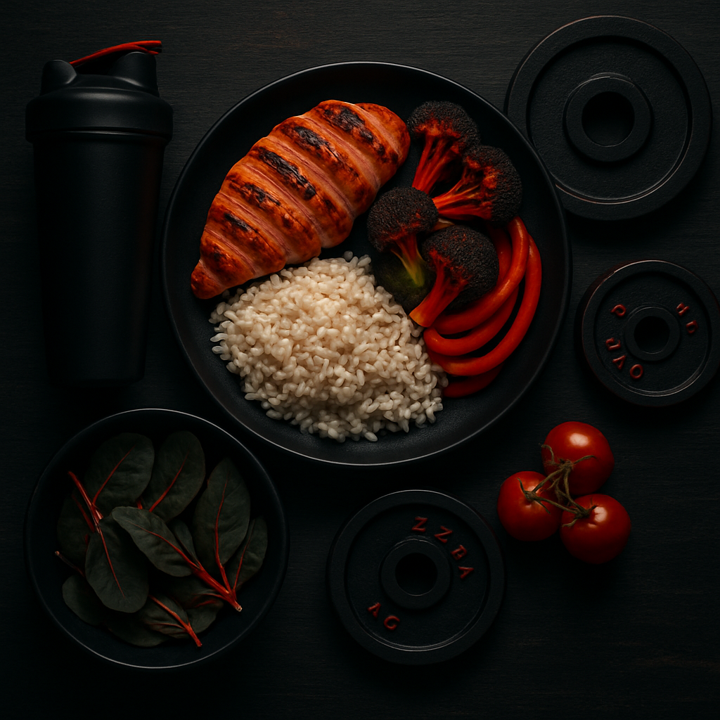
Ferritin & Iron Deficiency Evaluator
Evaluate serum ferritin status for endurance athletes (>50 mcg/L target) and compute gentle chelated iron protocols.
Launch Tool4. Why Athletes Lose Iron: Hemolysis, Sweating, and Hepcidin
Competitive athletes experience unique, accelerated rates of iron depletion driven by multiple concurrent biomechanical, physiological, and metabolic stressors that far exceed normal sedentary turnover. A prominent physical mechanism is foot-strike hemolysis, common in distance runners, triathletes, and field sport athletes. Repetitive mechanical shock waves transmitted through plantar capillary beds during foot strike crush circulating erythrocytes against skeletal structures, releasing free hemoglobin into plasma. This free hemoglobin is bound by haptoglobin and cleared by the reticuloendothelial system, but intense continuous training can saturate clearance pathways, leading to renal iron loss.
Perspiration represents another significant avenue of iron elimination during rigorous training. High-volume endurance exercise in temperate or hot climates elevates core temperature and stimulates heavy sweating, with sweat iron concentrations ranging between 0.3 and 1.0 mcg/mL. Over multi-hour training sessions or double-day practice regimens, cumulative dermal iron losses can exceed 1 to 2 mg per day. When combined with gastrointestinal micro-bleeding caused by transient splanchnic hypoperfusion during maximal efforts, daily iron losses can rapidly surpass dietary absorption rates.
The most potent regulatory mechanism of athletic iron depletion is the exercise-induced hepcidin surge. High-intensity muscular contractions, eccentric loading, and glycogen depletion trigger substantial intramuscular release of interleukin-6 (IL-6), a primary pro-inflammatory cytokine. Plasma IL-6 levels peak immediately following strenuous workouts and stimulate hepatic hepcidin gene transcription. Hepcidin concentrations peak between 3 and 6 hours post-exercise, closing intestinal ferroportin channels and completely halting dietary iron absorption during this critical metabolic window.
Female athletes encounter an additional layer of physiological vulnerability due to menstrual blood loss, which accounts for an average loss of 15 to 30 mg of elemental iron per cycle, and substantially more in athletes with menorrhagia. When heavy menstrual losses coincide with high-impact hemolysis, profuse sweating, and elevated exercise-induced hepcidin, maintaining iron equilibrium through standard dietary intakes becomes mathematically impossible without targeted nutritional planning.
5. Practical Dietetics: Heme and Non-Heme Iron in the Athlete's Diet
Optimizing athletic iron intake requires understanding the distinct physiological kinetics between dietary heme and non-heme iron sources. Heme iron, derived from the hemoglobin and myoglobin of animal tissues such as beef, liver, venison, dark poultry, and shellfish, exists in the reduced ferrous (Fe2+) state. It is absorbed intact through duodenal mucosal cells via specialized heme carrier protein 1 (HCP1) with a high bioavailability of 15% to 35%. Importantly, heme iron absorption remains largely immune to the inhibitory dietary compounds that commonly undermine plant-based iron bioavailability.
Conversely, non-heme iron constitutes the predominant form found in plant foods such as lentils, chickpeas, dark leafy greens, pumpkin seeds, oats, and fortified cereals. Non-heme iron exists primarily in the oxidized ferric (Fe3+) state, which precipitates as insoluble ferric hydroxide in the alkaline duodenal lumen unless reduced to the ferrous state by the brush-border enzyme duodenal cytochrome b (Dcytb) before transport by divalent metal transporter 1 (DMT1). Consequently, non-heme iron bioavailability is inherently lower, ranging between 2% and 8% in typical diets.
Dietary enhancers and inhibitors dramatically alter non-heme iron absorption. The co-ingestion of ascorbic acid (vitamin C) forms soluble iron-ascorbate chelates in the acidic gastric environment, increasing non-heme iron uptake up to threefold. Similarly, the "meat-protein factor" (cysteine-containing peptides from muscle tissue) significantly enhances non-heme absorption. In contrast, dietary phytates in unrefined grains, polyphenols and tannins in tea and coffee, and calcium in dairy products or supplements forcefully chelate iron, completely suppressing its duodenal uptake when consumed within the same meal window.
Athletes should strategically structure their meal timing to maximize absorption efficiency. High-iron meals or iron supplements should be scheduled away from caffeine- and dairy-rich foods, ideally paired with vitamin C sources such as citrus, bell peppers, or berries. Crucially, oral iron intake should be timed either first thing in the morning before training or at least 6 hours after strenuous workouts to circumvent the exercise-induced hepcidin spike that paralyzes duodenal iron transport.
6. Diagnostics: How to Correctly Read Ferritin and Iron Tests
Accurate clinical evaluation of iron status in athletic populations demands precise timing and a comprehensive panel of hematological biomarkers. The standard clinical gold standard for assessing storage iron is serum ferritin, where 1 mcg/L of circulating ferritin corresponds to approximately 8 to 10 mg of storage iron in healthy individuals. However, ferritin is also a positive acute-phase reactant synthesized in response to tissue inflammation and cytokine signaling. Following strenuous training, muscle damage can artificially elevate ferritin levels for 48 to 72 hours, producing false-negative results that mask true cellular iron depletion.
To ensure diagnostic accuracy, blood samples must be collected in a rested, fasted state at least 48 to 72 hours following the last exhaustive workout, and ideally tested alongside C-reactive protein (CRP) to rule out systemic inflammation. While general population guidelines define ferritin deficiency as <15 mcg/L, sports science consensus establishes that endurance and power athletes require ferritin levels above 35 mcg/L for baseline metabolic function, with an optimal athletic target of >50 mcg/L to sustain elite mitochondrial enzyme activity and erythropoietic drive.
A comprehensive athletic iron panel must also measure serum iron, total iron-binding capacity (TIBC), and transferrin saturation (TSAT), calculated as (Serum Iron / TIBC) * 100. Transferrin saturation reflects the immediate availability of iron for erythropoiesis; values below 16% to 20% indicate iron-restricted red blood cell production regardless of whether hemoglobin has yet declined. Soluble transferrin receptor (sTfR) assays provide an additional non-acute-phase marker that directly quantifies cellular iron demand even in the presence of exercise-induced systemic inflammation.
| Biomarker | Sedentary Reference Range | Athletic Optimal Target | Clinical Significance in Sports |
|---|---|---|---|
| Serum Ferritin | 15 – 150 mcg/L (F) / 20 – 300 (M) | > 50 mcg/L (both sexes) | Indicates total storage iron; values <35 mcg/L impair VO2 max and mitochondrial enzyme synthesis. |
| Transferrin Saturation (TSAT) | 20% – 50% | 25% – 45% | Reflects circulating iron availability; values <20% indicate iron-restricted erythropoiesis. |
| Hemoglobin (Hb) | 120 – 155 g/L (F) / 135 – 175 (M) | 130 – 150 g/L (F) / 145 – 165 (M) | Systemic oxygen carrying capacity; drops indicate clinical anemia requiring rapid medical intervention. |
| Soluble Transferrin Receptor (sTfR) | 1.0 – 2.5 mg/L | 1.0 – 2.0 mg/L | Direct marker of tissue iron demand; remains unaffected by acute exercise-induced inflammation. |
| C-Reactive Protein (CRP) | < 3.0 mg/L | < 1.0 mg/L | Inflammatory screen to verify that measured ferritin is not artificially elevated by tissue microtrauma. |
7. Scientific Research Analysis: The Impact of Iron Deficiency on VO2 Max and Endurance
A robust body of peer-reviewed sports science literature demonstrates that both frank anemia and non-anemic iron deficiency exert profound negative effects on athletic performance and physiological adaptation. Seminal investigations using treadmill ergometry and open-circuit spirometry confirm that even when hemoglobin concentrations remain stable, depleted iron stores (ferritin <20 mcg/L) reduce VO2 max by 5% to 10% and significantly shorten time-to-exhaustion at submaximal thresholds. This decrement stems directly from the downregulation of iron-dependent mitochondrial enzymes, specifically succinate dehydrogenase and cytochrome c oxidase.
Randomized double-blind placebo-controlled trials in elite female collegiate rowers, runners, and cross-country athletes have evaluated the effect of oral iron supplementation in non-anemic iron-deficient subjects. Athletes receiving daily elemental iron demonstrated statistically significant reductions in 2-km and 5-km time trial performance times compared to placebo controls. Concurrently, post-exercise blood lactate curves revealed that iron-replete athletes experienced significantly delayed lactate accumulation, reflecting superior oxidative phosphorylation efficiency and reduced reliance on compensatory anaerobic glycolysis.
Meta-analyses pooling data across hundreds of endurance competitors indicate that correcting iron deficiency without anemia improves gross energetic efficiency and lowers submaximal heart rates at equivalent power outputs. In cycling ergometer tests, iron-supplemented cohorts achieved higher peak power outputs and maintained elevated power at the ventilatory threshold. These findings refute the outdated dogma that only frank anemia impacts performance, establishing that cellular iron stores are critical determinants of energetic economy.
Furthermore, sports science research underscores the role of iron in central nervous system fatigue and cognitive executive function. Iron acts as an essential cofactor for tyrosine hydroxylase, the rate-limiting enzyme in dopamine synthesis. Athletes with suboptimal ferritin exhibit reduced reaction times, impaired motor coordination under fatigue, and degraded mood states. Restoring systemic iron homeostasis optimizes neurotransmitter turnover, reinforcing mental resilience during grueling endurance events.
8. Synergy: The Role of Vitamin C, Copper, and B12 in Iron Metabolism
Iron metabolism does not operate in biochemical isolation; rather, it functions within an intricate web of micronutrient synergies that govern its absorption, systemic transport, and utilization in erythropoiesis. Ascorbic acid (vitamin C) is the most potent exogenous enhancer of non-heme iron uptake. Beyond maintaining iron in the soluble Fe2+ state, vitamin C modulates cellular iron uptake by stimulating ferritin synthesis, preventing ferritin degradation, and inhibiting cellular iron efflux via autocrine mechanisms, making it an indispensable partner in nutritional iron strategies.
Copper plays a pivotal, non-negotiable role in iron mobilization and hemoglobin synthesis through the multicopper ferroxidases ceruloplasmin and hephaestin. Hephaestin, situated on the basolateral membrane of duodenal enterocytes, oxidizes newly absorbed Fe2+ into Fe3+, allowing it to bind apotransferrin for systemic distribution. Similarly, circulating ceruloplasmin oxidizes iron released from hepatic and macrophage stores. In states of subclinical copper deficiency, iron becomes trapped within cellular storage compartments, resulting in a refractory secondary microcytic anemia that fails to respond to oral iron therapy alone.
Vitamin A and its carotenoid precursors exert significant synergistic effects by forming soluble complexes with iron in the gastrointestinal tract, preventing phytate-mediated inhibition. Moreover, retinoic acid directly stimulates erythropoietin (EPO) gene transcription in renal fibroblasts and promotes the mobilization of hepatic iron stores into circulating transferrin. Clinical trials demonstrate that combined supplementation with vitamin A and iron resolves iron deficiency and elevates hemoglobin more rapidly than iron alone.
Finally, the water-soluble B vitamins, particularly cobalamin (vitamin B12) and folate (vitamin B9), are critical determinants of normal erythroid maturation. Vitamin B12 serves as a cofactor for methionine synthase, while folate supplies one-carbon units for thymidylate synthesis. A deficiency in either nutrient impairs red blood cell nuclear maturation, causing macrocytic megaloblastic changes that can coexist with or mask microcytic iron deficiency, leading to severe hematological dysfunction and delayed muscle oxygen delivery.
9. Common Mistakes, Myths, and How to Fix Them
Despite extensive scientific consensus, persistent misconceptions regarding iron management continue to compromise athlete health and performance. A prevalent and dangerous error is unmonitored, prophylactic mega-dosing of oral iron supplements without baseline blood testing. The human body lacks an active physiological mechanism for excreting excess iron; beyond controlled shedding of enterocytes, excess iron accumulates in the liver, myocardium, and endocrine tissues. Chronic iron overload promotes the Fenton reaction, generating highly cytotoxic hydroxyl free radicals that induce systemic lipid peroxidation, cardiac fibrosis, and tissue damage.
Another widespread mistake involves suboptimal supplement timing relative to exercise bouts. Many athletes ingest iron supplements with pre-workout or post-workout meals, directly colliding with the peak post-exercise hepcidin window (3 to 6 hours after strenuous training). Hepcidin degrades ferroportin and closes intestinal absorption gates, causing the majority of ingested iron to pass unabsorbed into the colon. There, unabsorbed iron alters the gut microbiome, triggers mucosal inflammation, and causes severe gastrointestinal distress, nausea, constipation, and abdominal cramping.
A third major error is consuming iron alongside powerful absorption inhibitors. Washing down iron tablets or iron-rich meals with coffee, green tea, dairy products, or multivitamin tablets containing high doses of calcium, magnesium, or zinc drastically suppresses iron uptake through competitive transport inhibition and chelation. Sports practitioners must educate athletes to space iron doses at least two hours apart from caffeine and dairy intake to ensure optimal duodenal bioavailability.
Evidence-based best practices mandate oral iron supplementation with gentle, high-bioavailability chelated forms such as ferrous bisglycinate or microencapsulated iron. Groundbreaking pharmacological research confirms that alternate-day iron dosing (taking iron once every 48 hours in the morning) yields equivalent or superior total iron absorption compared to daily dosing while triggering significantly lower secondary hepcidin spikes and causing dramatically fewer gastrointestinal side effects.
Interactive Apps & Calculators for Article
Empirical mathematical algorithms and scientific formulas for sports optimization
10. FAQ: Answers to Common Questions
- What is the optimal ferritin level for competitive endurance and strength athletes?
- While standard clinical laboratory reference ranges consider any ferritin level above 15 mcg/L normal, extensive sports science research demonstrates that athletes require a minimum ferritin concentration of 35 mcg/L to avoid subclinical performance impairment, with an optimal athletic target of >50 mcg/L. Maintaining ferritin above 50 mcg/L ensures maximal activity of iron-dependent mitochondrial enzymes (succinate dehydrogenase, cytochrome c oxidase) and sustains adequate erythropoietic drive during intensive training blocks.
- Why is oral iron supplementation often recommended on an alternate-day rather than daily schedule?
- When a dose of oral iron (60 to 100 mg of elemental iron) is ingested, the body responds by elevating hepatic hepcidin production for up to 24 hours as a protective mechanism against iron overload. Taking iron every single day means subsequent doses are ingested while hepcidin levels are still elevated, which severely impairs duodenal ferroportin and drastically reduces absorption. Alternate-day dosing (every 48 hours) allows hepcidin to return to baseline, resulting in significantly higher fractional absorption, reduced colonic iron residue, and minimal gastrointestinal discomfort.
- Can female athletes manage iron balance solely through a vegetarian or vegan diet?
- While it is theoretically possible, female athletes on plant-based diets face significant physiological hurdles. Plant foods contain only non-heme iron (Fe3+), which has a low baseline bioavailability (2% to 8%) and is heavily inhibited by phytates and polyphenols. To maintain adequate iron balance without supplements, plant-based athletes must deliberately consume 1.8 times the standard recommended dietary allowance, aggressively implement culinary soaking, sprouting, and fermentation to degrade phytates, and strictly pair every iron-rich meal with potent vitamin C sources while avoiding coffee, tea, and calcium during meals.
- How soon after intense workouts can athletes take iron without hepcidin interfering with absorption?
- Intense exercise stimulates muscular interleukin-6 release, triggering hepatic hepcidin production that peaks between 3 and 6 hours post-exercise and remains elevated for up to 12 hours. Consequently, athletes should avoid taking iron during the immediate post-workout window. The optimal timing window is either first thing in the morning in a rested state prior to strenuous training, or in the evening on low-intensity recovery days, always taken on an empty stomach with a source of vitamin C.
- When should an athlete consider intravenous (IV) iron therapy instead of oral supplementation?
- Intravenous iron therapy is a specialized medical intervention reserved for cases of severe clinical iron deficiency anemia where rapid hematological restoration is critical, cases where oral iron causes intolerable gastrointestinal side effects, or situations involving chronic gastrointestinal malabsorption (e.g., celiac disease, inflammatory bowel disease). Intravenous administration bypasses intestinal hepcidin blockades entirely, but it requires strict physician supervision due to potential risks of anaphylactoid reactions, hypophosphatemia, and transient oxidative stress.